
fbpx

Please wait...


BLOG

No more posts

Hard skin under one big toe is more than the other and it’s splitting like paper cuts.

When hard skin keeps coming back, look at why the skin is protecting itself

A common complaint we hear from patients is:

“I constantly get hard skin underneath my big toe. It splits and feels like little paper cuts. I use foot cream, file it and have the callus removed during pedicures, but it always comes back.”


When callus repeatedly develops in exactly the same place, simply removing the hard skin may not address the reason it is forming.

Callus is often the skin’s response to repeated pressure, shear and friction. The location and pattern of the callus can therefore provide useful clues about how forces are travelling through the foot.

A patient with painful, splitting callus

In this particular case, the patient had recurring hard skin underneath her big toe which frequently split, producing a painful sensation similar to paper cuts.

She had tried creams, regular filing and intensive callus removal at pedicure salons, but the problem continued to return.

She also had areas of callus beneath the ball of the foot, particularly underneath the second metatarsal head, as well as callus and a corn around the fifth metatarsal head.

She had also noticed that her feet seemed to be becoming broader.

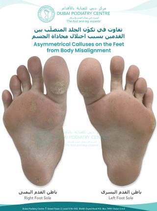
What was particularly interesting was the difference between her two feet.

The left foot had only a relatively mild amount of hard skin underneath the big toe and forefoot.

The right foot showed considerably more.

Right and left foot soles showing uneven calluses and hard skin beneath the big toes, more pronounced on the right foot

Why was one foot worse than the other?

When we see a significant difference between the right and left feet, we don’t just examine the skin.

We look higher up the body.

In this patient’s assessment, we identified a leg-length discrepancy associated with pelvic asymmetry. Her right side demonstrated a pelvic tilt and altered lower-limb position, with the right foot abducting as she walked.

Instead of progressing straight over the big toe, she was repeatedly rolling across the side of it.

This created increased pressure and shear around the plantar and side of the big toe and first metatarsal region.

The skin was therefore repeatedly being subjected to mechanical stress in the same location.

Removing the callus could make the skin temporarily smoother, but it did not remove the forces that were causing it to develop.

The callus in the middle of the foot told us something different

Not every area of callus on this patient’s foot had the same mechanical explanation.

She also had callus underneath the second metatarsal head and around the fifth metatarsal head.

During her examination, this corresponded with changes in the shape and loading of her forefoot and a reduction in the normal transverse arch across the front of the foot.

As the forefoot spreads and its loading pattern changes, individual metatarsal heads can become more prominent beneath the foot.

The second metatarsal head is a particularly common area of increased plantar pressure.

Between the metatarsal bones and the ground lies an important layer of specialised fibrofatty tissue that helps cushion the foot.

When excessive pressure and shear repeatedly occur over a prominent metatarsal head, the skin may respond by producing additional keratin.

This is what we recognise as callus.

A more concentrated area of pressure can produce a corn.

Callus isn’t necessarily the problem. It can be a clue.

This is an important distinction.

Callus is not simply “dead skin that needs to be removed”

Its distribution can act almost like a map of pressure across the foot.


A podiatrist will therefore look at questions such as:

  • Is the callus symmetrical between the two feet?
  • Is it underneath a particular metatarsal head?
  • Is it underneath or along one side of the big toe?
  • Is there a corn in the centre of the callus?
  • Has the forefoot become broader?
  • Is one arch lower than the other?
  • Does one foot pronate more than the other?
  • Is one leg functionally or structurally longer?
  • Is there pelvic asymmetry?
  • Is the patient’s walking pattern creating excessive pressure or shear?


The position of the callus can help us understand what may be happening mechanically above it.

Why do some calluses crack and feel like paper cuts?

When callus becomes thick, it is less flexible than normal skin.

Every time the toe bends, twists or pushes against the ground, that thickened skin is subjected to movement and shear.

Eventually tiny fissures can develop.

These cracks can feel surprisingly painful, sometimes exactly like a paper cut.

Applying moisturiser may improve the flexibility and condition of the skin, and careful reduction of excessive callus can provide considerable relief.

However, if abnormal pressure and shear remain, the skin may continue to rebuild the callus.

That is why some patients find themselves in a cycle:

Callus forms → callus is filed or removed → skin feels better → the same mechanical forces continue → callus returns


What about people with diabetes or neuropathy?

This is particularly important in patients who have reduced sensation in their feet.

A person with neuropathy may not feel excessive pressure, rubbing, a blister or a developing wound normally.

Callus can itself be an important warning sign of repetitive high pressure, and in a neuropathic foot, areas beneath callus may be at increased risk of tissue breakdown and ulceration.

For this reason, recurrent callus in a person with diabetes, neuropathy or reduced circulation should not simply be aggressively filed or treated with corn-removing acids without appropriate professional assessment.


Why we examine the whole body

At Dubai Podiatry Centre, when we see a recurrent pattern of callus, particularly when one foot is significantly worse than the other, we don’t only look at the piece of hard skin.

We assess the biomechanics producing the pressure.

This can include examining:

Pelvis → hips → knees → ankles → subtalar joints → arches → forefoot → toes

Human bodies are naturally asymmetrical. Small differences are extremely common and do not necessarily cause problems.

It is when that asymmetry produces excessive or repetitive loading that symptoms can begin to appear.

In this patient’s case, the difference between the right and left callus patterns was an important clue.


Why the right and left orthotics may be different

If biomechanical assessment indicates that orthotic therapy is appropriate, the objective is not simply to put identical arch supports underneath both feet.

The two sides of the body may require different corrections.

For example, one orthotic may require a different thickness, inclination or degree of support from the other. The medial arch support may differ, and the forefoot or transverse arch may require different accommodation or correction.

The prescription is based on the individual patient’s examination.

The aim is to redistribute plantar pressure and improve lower-limb and foot mechanics so that the skin is no longer repeatedly exposed to the same excessive forces.


How long does it take for the callus to stop returning?

Skin does not necessarily change immediately when biomechanics are altered.

Even after the source of excessive pressure has been reduced, the skin may continue producing protective keratin for some time.

In clinical practice, we may see the callus pattern gradually reduce over the following months as pressure and shear are better controlled. For some patients, noticeable changes may occur over approximately four to six months, although this varies considerably according to the individual, their footwear, activity, skin condition and underlying biomechanics.

The important point is that we are not simply asking:

“How do we remove this hard skin?”

We are asking:

“Why does this particular piece of skin keep needing to protect itself?”

Once we understand that, we can work on the underlying mechanical cause as well as treating the skin itself.

The takeaway

Recurring callus can be more than a cosmetic problem.

Its location, shape and whether it is symmetrical between the two feet can provide valuable information about pressure, friction, foot structure and walking mechanics.

If the same callus keeps returning despite creams, filing and pedicures, it may be time to stop treating only the surface and investigate what is happening underneath it.

Dubai Podiatry Centre
Looking beyond the callus to understand why it formed.


For more information or to book an appointment please call our clinic +971 4 3435390 or WhatsApp +971 50 3553024
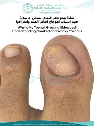


A big toenail that becomes white, lifted or separated along one side is often assumed to have a fungal infection. However, not every separated toenail is fungal.

In some patients, the underlying problem is mechanical and begins much further up the foot, at the alignment of the subtalar joint and ankle.

What causes the nail to separate?

When the foot pronates excessively, the subtalar joint and ankle lean inwards and the hallux, or big toe, can begin to internally rotate.

At the same time, the soft tissues and fibrofatty padding beneath the hallux are displaced laterally, towards the second toe.

Instead of remaining directly underneath the big toe, these tissues repeatedly move and press against the structures beneath and around the nail plate during standing and walking.

With thousands of steps each day, this repetitive movement can create an up-and-down and side-to-side mechanical force beneath the nail.

Over time, the nail plate can gradually be pulled away from the nail bed underneath. This produces a characteristic curved or U-shaped area of separation, particularly along the lateral border of the hallux nail.

This type of nail separation is known as onycholysis, but importantly, onycholysis describes the separation itself. It does not tell us what caused it.


An interesting left-versus-right comparison

This photographic example of the toes and feet provides an excellent example because the severity of the nail changes is different between the two feet.

On the left foot, the ankle/subtalar joint pronates approximately 30 degrees, and the lateral separation of the hallux nail plate is considerably greater.

On the right foot, the subtalar joint pronates approximately 12 degrees, and the lateral nail separation is noticeably less extensive.

The asymmetry is clinically interesting because the foot demonstrating the greater degree of pronation also demonstrates the greater degree of nail separation.

Rather than examining the toenail in isolation, we therefore need to examine what is happening to the whole foot, ankle and hallux during weight-bearing and walking.


Why can this be mistaken for fungal nail disease?

When air gets underneath a separated nail plate, the nail can appear white, yellowish or opaque. Debris may also accumulate beneath the separated portion.

Visually, this can resemble a fungal toenail.

However, treating a mechanically separated nail with antifungal treatment alone does not correct the forces repeatedly acting upon the nail.

Where fungal infection is suspected, appropriate clinical assessment and, when indicated, laboratory testing can help distinguish fungal nail disease from other causes of nail dystrophy and onycholysis.


The relationship with ankle alignment

In our clinical observations at Dubai Podiatry Centre, we frequently begin to see secondary foot and nail changes as subtalar joint pronation increases.

When inward deviation becomes more pronounced, mechanical stress upon the hallux can increase. With greater degrees of pronation, we may see progressively wider and deeper lateral nail separation together with increasing rotation or displacement of the hallux.

At very significant degrees of pronation, other structural changes, including hallux valgus, may also coexist.

These degree measurements should not be interpreted as universal diagnostic thresholds. Every patient is assessed individually because foot structure, joint mobility, footwear, activity, gait and other medical or dermatological factors can also influence the appearance of the toenail.

Treatment must address the cause, not simply the nail

If the separation is predominantly mechanical, our objective is to reduce the abnormal forces acting upon the hallux long enough for a healthier nail to grow forward while remaining attached to the nail bed.


Depending upon the individual assessment, treatment may include:

  • Corrective orthotics designed to improve subtalar joint alignment and reduce excessive pronation
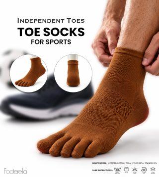
  • Toe socks to encourage the toes to remain separated and in a more neutral position
  • Silicone toe liners or separators between the toes where appropriate to assist alignment
  • Footwear with an accommodating toe box, including suitable barefoot-style footwear where clinically appropriate, so that the toes are not compressed or displaced by the shoe

The important principle is consistency.

If a corrective device is worn for only one hour each day but the foot spends the remaining weight-bearing hours returning to its previous position, the mechanical forces responsible for the problem continue.

The objective is to maintain improved foot and toe alignment for as much of the patient’s weight-bearing day as reasonably possible.


How long does the nail take to recover?

Toenails grow slowly.

The recovery period therefore depends partly upon how far backwards the existing separation extends. The damaged portion cannot simply be pushed back onto the nail bed. Instead, we need to reduce the causative mechanical stress and allow healthier nail to gradually grow forward.

For some patients, visible improvement may occur over several months. A mechanically separated area may require approximately four to six months or longer to grow forward sufficiently, depending upon the depth of separation, individual nail-growth rate and how effectively the underlying mechanical problem is controlled.

Look beyond the toenail

A separated toenail does not always mean a fungal toenail.

Sometimes the toenail is simply showing us the end result of a biomechanical problem occurring elsewhere in the foot.

By examining the nail together with the hallux position, plantar soft tissues, subtalar joint, ankle alignment, gait and footwear, we can investigate why the nail is separating rather than treating its appearance alone.

Michelle Duffy Champlin
B.Sc. (Podiatry), M.Ch.S., S.R.Ch. (UK)
Chief Podiatrist
Dubai Podiatry Centre


For more information or to book an appointment please call our clinic +971 4 3435390 or WhatsApp +971 50 3553024


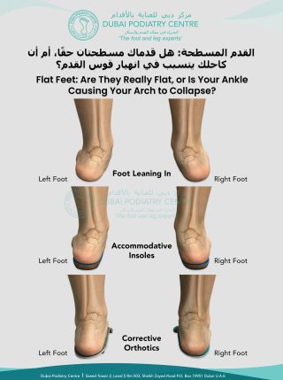
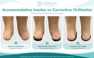
Comparison of inward-leaning ankles without support, with accommodative insoles, and with corrective orthotics


Do your ankles roll inward when you stand or walk? You may have been advised to try insoles, but different types of foot support serve different purposes. Understanding the difference can help you ask the right questions during a podiatry assessment.

What Are Accommodative Insoles?

Accommodative insoles are generally soft and cushioned. They are designed to support the foot and relieve pressure on sensitive areas. They may improve comfort for people with tender soles or painful calluses, but cushioning alone does not necessarily address abnormal foot alignment.

What Are Corrective Orthotics?

Corrective, or functional, orthotics are designed to influence how the foot moves inside the shoe. For some people with flat feet or overpronation, a prescribed orthotic may help manage excessive inward rolling, support foot position and redistribute pressure while walking.

The image shows inward-leaning ankles alongside accommodative insoles and corrective orthotics. It illustrates the difference between cushioning the foot and providing support intended to guide its movement.

Which Type of Foot Support Is Right for You?

The choice depends on your symptoms, foot structure, walking pattern and footwear. A podiatrist can assess how your feet and ankles function before recommending accommodative insoles, custom corrective orthotics or another approach.

If you experience foot pain, heel pain, arch discomfort or recurring calluses, book a biomechanical foot assessment at Dubai Podiatry Centre. We will evaluate your foot alignment and walking mechanics to recommend support suited to your needs.


For more information or to book an appointment please call our clinic +971 4 3435390 or WhatsApp +971 50 3553024


Many people become frustrated when trying to treat a plantar wart. They may have tried over-the-counter salicylic acid, cryotherapy (freezing), or various home remedies, only to find that the wart stubbornly remains.

The reason is simple: not all warts behave in the same way.


Why Warts on the Feet Are Different

A wart on the back of the hand or another area of the body often appears as a small raised lump. This is because the skin in these areas is relatively thin and flexible, allowing the wart to grow outward.

The sole of the foot is completely different.

The skin on the bottom of the foot is much thicker and stronger because it is designed to withstand the pressure of standing and walking. Instead of growing outward, a plantar wart is compressed by body weight and becomes trapped beneath the thick outer layers of skin.

As a result, much of the wart lies within the skin rather than sitting prominently on top of it. This is why plantar warts often appear relatively flat while extending deeper into the skin layers.


Why Some Treatments Work Better on Hand Warts Than Plantar Warts

Because common warts on the hands are more exposed, superficial treatments such as:

  • Salicylic acid
  • Cryotherapy (liquid nitrogen freezing)
  • Occlusion methods

can often work very well.

However, plantar warts are protected by the thick skin of the sole. Treatments that work primarily on the surface may struggle to reach the full extent of the infected tissue beneath.

This is one reason why treatment of plantar warts can be slower and sometimes less predictable than treatment of warts elsewhere on the body.


Our Approach at Dubai Podiatry Centre

At Dubai Podiatry Centre, our goal is to make the wart more accessible to the body’s natural healing mechanisms.

Rather than aggressively destroying tissue, we focus on improving the quality and softness of the overlying skin. As the thick layers gradually become softer and thinner, the body is better able to recognise and respond to the viral infection.

The tiny black dots often seen within a plantar wart are small blood vessels that have become altered by the wart tissue. These blood vessels are part of the body’s normal circulation and immune surveillance system.


Why We Use Vitamin A

Vitamin A helps regulate normal skin cell turnover and supports healthy skin function.

We recommend applying Vitamin A cream to the plantar wart each evening and covering it with a non-absorbent dressing overnight.

Treatment is usually continued for approximately three skin cycles.

A skin cycle lasts around 28 days, meaning treatment is typically continued for 12–14 weeks.

During this period, the thick overlying skin gradually softens and normalises, allowing the body’s immune system to recognise and clear the viral tissue more effectively.

Many patients notice significant improvement during this timeframe.


What Is Dermojet Treatment?

For stubborn or longstanding plantar warts, we often combine Vitamin A therapy with Dermojet treatment.

The Dermojet uses a high-pressure microjet of local anaesthetic delivered through an opening smaller than a conventional needle.

The medication passes through the skin and disperses within the deeper tissue layers without the need for a traditional injection.

This creates a controlled healing response within the affected tissue while avoiding significant damage to the skin surface.

As healing occurs, the body’s immune system is stimulated within the wart tissue, helping to clear the virus.

In our clinical experience, the combination of Vitamin A therapy and Dermojet treatment provides some of the most successful outcomes for difficult plantar warts.


Why We Do Not Routinely Cut Out Plantar Warts

Many people assume that simply cutting out a plantar wart would solve the problem.

Unfortunately, plantar warts often extend beyond what is visible on the surface.

To completely remove all affected tissue surgically, a large and deep section of skin may need to be excised.

On the sole of the foot, this can result in:

  • Significant scar tissue formation
  • Persistent discomfort when walking
  • Hard callus formation
  • Long-term pressure-related problems

For these reasons, surgical excision is generally not our preferred treatment option.


Why We Do Not Routinely Use Cryotherapy on the Sole of the Foot

Cryotherapy can be effective for some types of warts, particularly those located on thinner skin.

However, on the sole of the foot it often requires multiple treatments and can be quite painful.

In our experience, the success rate is often lower than patients expect, particularly for deeply embedded plantar warts.

We are also cautious when considering cryotherapy around the toes because freezing treatments affect local blood circulation and can occasionally cause unnecessary tissue injury.


Our Recommended Treatment Plan

For most plantar warts, we recommend:

1. Daily Vitamin A application.
2. Occlusion with a non-absorbent dressing overnight.
3. Treatment continued for approximately 12–14 weeks.
4. Dermojet therapy for persistent or resistant lesions.

By helping the skin return to a healthier state and stimulating the body’s natural healing response, many plantar warts can be cleared without aggressive surgery or repeated freezing treatments.

If you are struggling with a plantar wart that simply won’t go away, a consultation with a podiatrist can help determine the most appropriate treatment plan for your particular case.


For more information or to book an appointment please call our clinic +971 4 3435390 or WhatsApp +971 50 3553024


When we think about the arches of our feet, most people immediately picture the large arch running along the inside of the foot.

But there is another, much smaller arch at the front of the foot that we pay very close attention to during a biomechanical assessment at Dubai Podiatry Centre.

It is called the transverse arch.


What is the transverse arch?

The front part of your foot is called the forefoot. It includes the toes and the metatarsal bones, which are the long bones immediately behind your toes.

Across the forefoot, just behind the toes where they connect with the foot, is the transverse arch.

Rather than running from the heel towards the toes like the more familiar medial arch, the transverse arch runs across the foot from side to side.

It is a relatively small arch, but biomechanically it can be extremely important.


What happens when the transverse arch starts to drop?

Over time, the forefoot can begin to spread and the transverse arch can become lowered.

We sometimes describe this to patients as the metatarsal bones beginning to “avalanche” forwards and downwards.

Although this change is occurring in a relatively small area at the front of the foot, it can influence the way the rest of the foot functions.

The metatarsal bones do not work independently from the remainder of the foot. They form part of the interconnected structure of the foot and contribute to its stability and function during standing and walking.

As the forefoot begins to change position, we may also see changes occurring through the larger medial arch, the midfoot and eventually the mechanics around the ankle.

This is why we never look at the transverse arch in isolation.


3D postural assessment showing how a collapsed forefoot transverse arch may affect standing posture and spinal alignment


Are you beginning to look hunched forwards?

One of the observations that can prompt us to investigate the forefoot is a change in a person’s stature.

Sometimes a patient, or their family, will say that they are beginning to “look hunchbacked” or that they appear to be leaning forwards.

The head may be sitting further forwards, the shoulders may appear more rounded and the person can develop an increasingly stooped appearance.

There are, of course, many different reasons why somebody may develop a forward head position or stooped posture. It should never automatically be assumed that it is coming from the feet.

However, it is one of the physical signs that makes us look more carefully at the person’s overall biomechanics.

We look at the person from the side and assess their stature. We look at the position of the head and shoulders. We observe how they stand and walk.

Then we investigate what is happening underneath them.

In particular, we examine the sole of the foot and the forefoot to see what has happened to the transverse arch.

If that small arch has dropped and the metatarsal bones have moved forwards and downwards, it may be contributing to a chain of biomechanical changes occurring further up the body.

Your feet form the foundation of your posture


The feet provide the foundation for the body when we stand and walk.

Changes in that foundation can sometimes contribute to compensations further up the biomechanical chain.

This is why a person’s posture can give us clues about what we should investigate in their feet.

A forward head, rounded shoulders, a stooped stance or a tendency to shuffle when walking does not diagnose a foot problem. However, these observations can be a reason to include the feet in the assessment rather than concentrating only on the upper body.

Sometimes the clue is visible at the shoulders and head, while part of the biomechanical problem may be much further down, at the sole of the foot.


Why we look for problems early

One of the reasons we pay such close attention to the transverse arch is that it can give us an opportunity to identify biomechanical changes relatively early.

If the transverse arch has started to lower but the remainder of the foot is still relatively well controlled, treatment can be much simpler.

Where appropriate, a custom corrective orthotic can be designed to support and lift the transverse arch and improve the mechanical stability of the forefoot.

The objective is not simply to put something soft underneath a painful area. It is to address the position and function of the forefoot as part of the mechanics of the entire foot.


What if the whole foot has already started to collapse?

More advanced biomechanical changes can also be managed. It simply requires a different approach.

If changes have progressed through the forefoot, midfoot and ankle, attempting to correct everything at once may not be appropriate or comfortable.

Instead, orthotic therapy may need to be carried out step by step and stage by stage.

We may initially need to address the forefoot and transverse arch. As the foot adapts, we can reassess the midfoot, medial arch, rearfoot, heel and ankle mechanics.

This process naturally takes longer than treating an early biomechanical change.

A small arch can give us a big clue

The transverse arch is a relatively small structure, but it can tell us a great deal about what is happening throughout the foot.

This is why we don’t assess feet simply by asking, “Where does it hurt?”

We look at the toes, forefoot, midfoot, heel and ankle. We look at how you stand. We look at the position of your shoulders and head. We watch how you walk.

If somebody tells you that you are beginning to look more hunched over, or you notice that your head is increasingly sitting forwards and your shoulders are becoming rounded, there can be many possible explanations.

But don’t forget to look down.

A biomechanical assessment of the feet, and particularly that small transverse arch underneath the forefoot, may provide another important piece of the puzzle.

The earlier we identify a change in foot biomechanics, the easier it may be to manage before it progresses further.


Dubai Podiatry Centre
Biomechanical Foot Assessment and Custom Orthotic Therapy


For more information or to book an appointment please call our clinic +971 4 3435390 or WhatsApp +971 50 3553024


A very small area of sudden, intense and highly localised pain in the sole of the foot can sometimes be caused by a foreign object embedded in the skin.

Because we put our full body weight through our feet, even something incredibly small—a tiny splinter, hair or fragment of glass—can become surprisingly painful. Patients will often describe being able to point to one exact spot that feels extremely sharp or painful when they stand or walk.


Even a Hair Can Become a Foreign Body

One of the more unusual foreign bodies we occasionally encounter is human hair.

For example, when someone shaves in the shower, freshly cut hairs can have extremely sharp ends. At the same time, warm water temporarily softens the outer layers of skin on the feet.

A short, stiff piece of freshly cut hair can occasionally penetrate this softened skin. With continued walking and pressure, it may gradually work deeper into the skin.

This can also occur with cat or dog hairs, particularly short, coarse hairs that can behave almost like tiny splinters.

Initially, the person may notice very little. However, as the foreign material penetrates more deeply and the surrounding tissue becomes inflamed, a very small area can become disproportionately painful.


Glass splinter removed from the sole of the foot following assessment and careful debridement at Dubai Podiatry Centre


Why Does It Become So Painful?


The body recognises an embedded object as something that does not belong there and produces an inflammatory response around it. Occasionally, a small collection of pus may develop around the foreign material.

In other areas of the body, this may eventually drain spontaneously and allow the foreign body to escape.

The sole of the foot, however, has particularly thick, strong skin designed to withstand enormous amounts of pressure and friction. This can make spontaneous drainage more difficult.

Instead, the foreign object may remain trapped while every step continues to push pressure through the affected area.

The result can be intense pain concentrated within a surprisingly tiny area.


Common Foreign Objects We Find in Feet

Foreign bodies can enter the feet in many everyday situations. These include:

  • Tiny wooden splinters from gardening, decking or wooden floors
  • Small fragments of broken glass
  • Human hair following shaving
  • Coarse cat or dog hairs
  • Thorns and plant material
  • Small fragments picked up while walking barefoot outdoors
  • Splinters from harbour or marina decking
  • Sea urchin spines

Walking barefoot around swimming pools, gardens, beaches, marinas and decking areas can increase the opportunity for tiny objects to enter the skin.

Look Closely at the Area

Sometimes there is very little to see from the surface.

You may notice a tiny dark or discoloured area beneath the skin. Occasionally patients describe seeing a greenish or grey shadow, although colour alone cannot confirm that a foreign body is present.

One simple trick is to take a close-up photograph with your mobile phone in good lighting and enlarge the image. Modern phone cameras can sometimes reveal a tiny entry point or object that is difficult to see with the naked eye.

However, if you cannot easily see and grasp the object at the surface, repeatedly digging into the foot at home can make matters worse.

Sea Urchin Spines

Sea urchin injuries are usually much easier for the patient to recognise because they occur immediately after standing on or brushing against a sea urchin.

The difficulty is that sea urchin spines are fragile and can break, leaving fragments embedded beneath the skin.

At Dubai Podiatry Centre, we use careful podiatric techniques to access suitable superficial retained fragments and remove them while trying to avoid breaking the spine or driving it further into the tissues.

Deeply embedded fragments, significant swelling, persistent pain or suspected involvement of deeper structures may require further medical assessment or imaging.

Don’t Keep Digging at the Foot

One of the most important pieces of advice is not to repeatedly cut, squeeze or dig into the sole of the foot yourself.

If a foreign body is clearly protruding from the surface and can be removed easily, that may be straightforward. But when an object has disappeared beneath the skin, trying to find it with needles, blades or other instruments can push it deeper, introduce bacteria and cause additional tissue damage.

If you have a very small, intensely painful area on the foot—particularly after walking barefoot—and you cannot see what is causing it, a podiatrist can examine the area carefully.


When Should You Seek Medical Attention?


Seek professional assessment if the pain is increasing, you cannot comfortably walk, the object appears deeply embedded, or you develop redness, warmth, swelling, pus, bleeding or spreading inflammation.

People with diabetes, poor circulation, reduced sensation in the feet or impaired immunity should avoid attempting to remove embedded foreign bodies themselves and should seek professional assessment promptly.

Sometimes the tiniest object can cause an extraordinary amount of pain in the foot. Finding and safely removing the cause can provide enormous relief.

Dubai Podiatry Centre
Dubai, UAE
Specialist assessment and treatment of foot and lower-limb conditions.


For more information or to book an appointment please call our clinic +971 4 3435390 or WhatsApp +971 50 3553024


Plantar fasciitis is one of the most common causes of heel and arch pain. A typical symptom is pain underneath the heel when you first get out of bed in the morning, or when you stand up after sitting at your desk, driving or relaxing for a period of time.

The plantar fascia is a strong band of connective tissue running along the sole of the foot from the heel towards the forefoot. It plays an important role in supporting the arch and stabilising the foot during standing and walking.

When the plantar fascia is repeatedly placed under excessive tensile stress, particularly where it attaches to the calcaneus (heel bone), the area can become painful and irritated.

Importantly, plantar fasciitis is not always simply a problem with the plantar fascia itself. Foot and lower-limb biomechanics can contribute to excessive loading of the fascia. This is why successful treatment should aim not only to settle the painful area, but also to identify and reduce the mechanical stresses that are causing it.

Here are some simple measures that can be carried out at home.


1. Contrast Foot Baths – Warm and Cool Water Therapy

Contrast bathing can be a useful part of a home-care routine.

Prepare two containers deep enough for the water to reach approximately midway up the lower legs.

Warm bath: approximately 40–42°C
Cool bath: approximately 22°C

Place both feet and lower legs into the warm water for approximately one minute.

Then transfer them into the cool water for one minute.

Continue alternating between the warm and cool water, repeating the process approximately five times. The complete treatment should take around 10–12 minutes.

The change in temperature produces alternating warming and cooling of the tissues and changes in local blood flow. Many patients find this soothing, particularly towards the end of the day.

We generally recommend doing contrast bathing in the latter part of the day rather than immediately before going to bed, as some people find the treatment quite stimulating.

Always check the temperature carefully before immersing your feet. Contrast bathing may not be appropriate if you have reduced sensation, neuropathy, vascular disease, diabetes-related foot complications or another condition affecting your ability to accurately feel temperature.


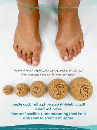
2. Massage the Plantar Fascia with Cork Balls

Cork balls are an excellent and simple way of gently massaging the sole of the foot while sitting at home or at your desk.

Different-sized cork balls can be used for different areas of the foot.

With a small cork ball, place the ball underneath the heel while seated and gently rest the foot over it. Make small circular movements around the plantar heel, alternating between clockwise and anticlockwise movements.

A medium or larger cork ball can be placed underneath the arch. Gently roll the foot backwards and forwards over the ball to massage the plantar fascia and surrounding soft tissues.

This is particularly useful if you spend long periods sitting.

One of the characteristic symptoms of plantar fasciitis is pain when standing after rest. Gentle movement and massage before standing can help prepare the foot for loading again.

The massage should feel comfortable rather than painful. Aggressive rolling directly over an acutely painful heel is unnecessary and can aggravate already sensitive tissue.


3. Plantar Fascia Support Tape

Traditional plantar fascia taping often involves applying several individual strips of tape underneath and around the foot. Although this can be effective, it can also be cumbersome and difficult for patients to apply themselves.

A purpose-designed single plantar fascia support tape can be a much simpler home option.

The tape is only a few millimetres thick and can provide gentle padding underneath the foot while helping to reduce some of the tensile load placed upon the plantar fascia.

It is generally worn during the daytime, when the foot is weight-bearing and the plantar fascia is repeatedly being loaded.

Taping is not intended to permanently correct the underlying biomechanics. Instead, think of it as temporary mechanical assistance while the irritated tissue is recovering.


4. Support the Arch and Correct Excessive Foot Movement

If plantar fasciitis continues to return, it is important to look at why the plantar fascia is being overloaded.

The plantar fascia does not work independently from the rest of the foot and leg.

For example, excessive movement through the subtalar joint and lowering of the arch can increase the mechanical demands placed upon the plantar fascia. Changes in the forefoot and transverse arch may also affect the way the foot loads during walking.

When the foot is not functioning efficiently, the plantar fascia may be subjected to increased tension as it contributes to supporting and stabilising the foot.

This is why a suitable supportive insole or individually prescribed corrective foot orthotic can be extremely beneficial for some patients.

The purpose is not simply to cushion the painful heel. A properly selected device should address the mechanical forces contributing to the problem and reduce unnecessary tensile stress through the plantar fascia.

For patients with significant biomechanical abnormalities, a professional assessment is advisable rather than simply purchasing a very soft heel cushion.


5. Give Your Toes Enough Space

Footwear is another important part of plantar fasciitis management.

The toes are designed to spread and function as part of the foot’s natural support system. Constantly squeezing them together inside narrow or pointed footwear can interfere with normal forefoot mechanics.

We often recommend toe socks combined, where appropriate, with very soft silicone toe aligners to allow the toes to sit in a more natural position.

These can be combined with an orthotic incorporating appropriate arch and transverse-arch support.

The shoe itself should have sufficient width through the toe box to allow the toes to open rather than compressing them together.

For some patients, a wider, foot-shaped or barefoot-style toe box can be very helpful. However, footwear needs to be selected according to the individual foot and the stage of recovery; a sudden transition into very flexible or minimally cushioned footwear may not be appropriate during an acutely painful episode.


Dubai Podiatry Centre infographic showing home treatment options for plantar fasciitis, including cork-ball massage, plantar fascia support tape, corrective orthotics, toe aligners and toe socks


How Long Does Plantar Fasciitis Take to Heal?

This is one of the most important things to understand.

Plantar fasciitis rarely disappears overnight.

The plantar fascia is a very strong, highly loaded structure, and the attachment at the heel is repeatedly stressed every time you stand and walk.

Even when the underlying mechanical problem is being corrected and the symptoms begin to improve, it can take several months for the area to settle fully. In many of the patients we treat, we advise allowing approximately three to four months for progressive recovery, although individual healing times vary.

The key is consistency.

Contrast bathing, gentle cork-ball massage, plantar fascia support taping, appropriate footwear and correctly prescribed arch support can all work together to reduce stress on the painful area while the tissues recover.

Don’t Just Treat the Heel – Find Out Why It Hurts

Perhaps the most important message is that plantar fasciitis should not always be viewed simply as an isolated painful heel.

Persistent plantar fascia pain can be a sign that the foot is compensating for a wider biomechanical issue.

The position and movement of the forefoot, arch, subtalar joint, ankle, knee, pelvis and hip can all influence how forces are transmitted through the foot.

If your plantar fasciitis keeps returning, is becoming progressively worse, or has not improved despite appropriate home treatment, it is worth having your walking pattern and lower-limb biomechanics professionally assessed.

At Dubai Podiatry Centre, we assess the foot as part of the complete lower-limb mechanical system. By identifying why the plantar fascia is being overloaded, treatment can be directed towards both relieving the painful tissue and reducing the mechanical stresses that contributed to the problem in the first place.

Dubai Podiatry Centre
Helping your feet function better from the ground up.

This article provides general educational information and is not a substitute for individual medical assessment. Persistent or severe heel pain, swelling, neurological symptoms, inability to bear weight, or pain following an injury should be professionally assessed.


For more information or to book an appointment please call our clinic +971 4 3435390 or WhatsApp +971 50 3553024


Flat feet in children are common, but parents may become concerned when a child’s feet or ankles roll inward while standing or walking. This inward movement is known as overpronation and may affect foot posture, ankle stability, balance and walking comfort.

At Dubai Podiatry Centre, our podiatrists assess children’s flat feet and walking patterns to determine whether additional support, such as custom-made orthotics, may be beneficial.


Child’s flat feet showing overpronation and improved neutral foot alignment with custom orthotics in Dubai

What Are Flat Feet in Children?

Flat feet occur when the arches of the feet appear lowered or make increased contact with the ground. In some children, the heels and ankles may also lean inward, causing the feet to overpronate.

Although flat feet do not always cause problems, a podiatry assessment may be recommended if your child experiences:

  • Foot, heel, ankle or knee pain
  • Tired or aching legs
  • Frequent tripping or poor balance
  • Difficulty walking or participating in sports
  • Uneven or excessive shoe wear
  • Ankles that visibly roll inward
  • Changes in posture or walking pattern

Recognising these signs early allows the child’s foot posture, movement and support requirements to be professionally evaluated.

How Overpronation Can Affect Foot Alignment

When a child’s feet overpronate, the heels and ankles lean inward instead of remaining in a more neutral position. This may change how pressure is distributed across the feet and influence the alignment of the lower limbs.

The comparison image demonstrates the rear view of pronating feet and the improved neutral alignment achieved while standing on corrective orthotics. The orthotics provide support beneath the feet and help stabilise the heels and ankles.

Custom Orthotics for Children’s Flat Feet

Custom orthotics are individually made to match the shape, posture and biomechanical needs of each child’s feet. Unlike standard insoles, they are created following a professional foot and walking assessment.

Corrective orthotics may help to:

  • Support the arches of the feet
  • Reduce excessive inward rolling
  • Stabilise the heels and ankles
  • Improve foot and lower-limb alignment
  • Distribute pressure more evenly
  • Support balance and walking comfort
  • Reduce strain on the feet and legs

Every child’s feet are different, which is why orthotics should be designed according to their individual assessment rather than selected as a one-size-fits-all solution.

Children’s Biomechanical Foot Assessment in Dubai

At Dubai Podiatry Centre, a children’s biomechanical assessment examines foot posture, ankle alignment, lower-limb movement and walking pattern. The podiatrist will determine whether custom orthotics are appropriate and explain the recommended treatment plan.

Regular reviews may also be advised to monitor the child’s progress, comfort and changing needs as they grow.

Book a Children’s Flat Feet Assessment in Dubai

If your child’s feet roll inward or you have concerns about flat feet, foot pain, balance or walking, professional assessment can help identify their individual support requirements.

Book a children’s flat feet and overpronation assessment with Dubai Podiatry Centre on Sheikh Zayed Road. Our podiatrists provide personalised foot assessments and custom orthotics for children in Dubai.


For more information or to book an appointment please call our clinic +971 4 3435390 or WhatsApp +971 50 3553024


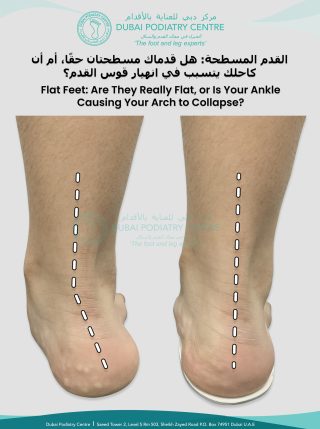
Flat feet can affect more than just the feet. When the arches collapse excessively during standing or walking, the feet may roll inwards—a movement known as overpronation. This can alter the position of the heels and affect the alignment of the ankles, knees and hips.

Over time, poor foot alignment may place additional strain on the muscles, tendons and joints throughout the lower limbs.


Signs and Symptoms of Flat Feet

Not everyone with flat feet requires treatment. However, adults and children whose flat feet cause pain or affect their mobility may experience:

  • Tired or aching feet
  • Heel, arch or ankle pain
  • Knee or hip discomfort
  • Reduced walking endurance
  • Difficulty standing for long periods
  • Uneven shoe wear
  • Changes in walking pattern

A professional foot and gait assessment can help determine whether flat feet or overpronation are contributing to these symptoms.

Corrective Orthotics for Flat Feet

Not all insoles provide the same type of support. Cushioned insoles are primarily designed to improve comfort and redistribute pressure. Corrective orthotics are designed to control excessive foot movement, support the position of the heels and improve lower-limb alignment.

When clinically appropriate, custom-made corrective orthotics may help reduce excessive strain on the feet and legs, improve stability and make standing, walking and daily activities more comfortable.

Children’s and adults’ feet have different requirements. For this reason, corrective orthotics should be individually prescribed following a detailed assessment rather than selected as a one-size-fits-all solution.

Biomechanical Assessment at Dubai Podiatry Centre

At Dubai Podiatry Centre, flat feet treatment begins with a comprehensive biomechanical assessment. Our podiatrist evaluates:

  • Foot posture and arch position
  • Heel and ankle alignment
  • Walking pattern and gait
  • Muscle function
  • Joint movement
  • Lower-limb biomechanics

The assessment helps identify how the feet are functioning and whether custom-made corrective orthotics are suitable for you or your child.

Book a biomechanical assessment at Dubai Podiatry Centre to learn more about flat feet, overpronation and corrective orthotic treatment.


For more information or to book an appointment please call our clinic +971 4 3435390 or WhatsApp +971 50 3553024


Choosing the right shoes is especially important for children who wear custom foot orthotics. Orthotic-friendly shoes should provide enough space for the devices while keeping the feet secure, comfortable and properly supported throughout the day.

At Dubai Podiatry Centre, we help parents choose suitable children’s footwear for custom orthotics, flat feet, overpronation and other foot conditions.

Why Are Orthotic-Friendly Shoes Important?

Custom orthotics are designed to support a child’s feet, improve foot function and distribute pressure more evenly. However, the orthotics must fit correctly inside the shoes to work as intended.

Shoes that are too narrow, shallow, soft or unstable may cause the orthotics to move, lift the heel or create pressure and discomfort. The best shoes for kids wearing orthotics should securely accommodate the devices without restricting the feet.

What to Look for in Children’s Shoes for Orthotics

When shopping for supportive kids’ shoes, look for the following features:

Removable Insoles

Shoes with removable insoles create additional space for custom orthotics. The original insoles should normally be removed before inserting the orthotic devices.

A Wide Toe Box

A wide, rounded toe box allows the toes to move naturally without rubbing or being compressed. This is particularly important as children’s feet continue to grow.

Adequate Width and Depth

The shoes must be deep and wide enough to accommodate the orthotics without making the fit too tight or pushing the feet upwards.

A Firm Heel Counter

A supportive heel counter helps hold the heel securely and reduces unwanted movement inside the shoe. This can provide additional stability for children with flat feet or overpronation.

Adjustable Fastenings

Laces, straps or adjustable closures help keep the feet and orthotics securely positioned. They also allow the fit to be adjusted as needed.

A Supportive Sole

Choose shoes with a stable sole that bends naturally near the toes rather than twisting or folding easily through the middle.

Lightweight and Durable Materials

Children need footwear that is comfortable for daily movement while being strong enough for school, sports and everyday activities.

Shoes for Children With Flat Feet and Overpronation

Children with flat feet or overpronation may require extra support to improve stability and reduce excessive inward rolling of the feet. Combining professionally prescribed custom orthotics with appropriately fitted supportive shoes can improve walking comfort and help the orthotics work effectively.

Every child’s feet are different. The best shoe is not simply the most expensive or popular option—it is the shoe that fits the child’s feet and orthotics correctly.

Tips for Finding the Correct Fit

Always take your child’s orthotics when shopping for new shoes. Insert the devices and ask your child to walk in the shoes before making a decision.

Check that:

  • The orthotics sit flat and securely inside the shoes.
  • The heels do not slip while walking.
  • The toes have enough room to move comfortably.
  • There are no areas of rubbing or pressure.
  • The shoes feel secure without being tight.
  • Your child can walk naturally and comfortably.

Do not automatically buy a larger shoe size to create space for the orthotics. A shoe that is too large may allow the feet and orthotics to move excessively. Instead, choose footwear with the correct width, depth and internal shape.

Expert Footwear Advice in Dubai

If your child has flat feet, overpronation, foot discomfort or difficulty finding shoes that fit their orthotics, a professional foot assessment can help.

Watch our video to discover the best supportive shoes for kids wearing orthotics, recommended by Michelle Duffy Champlin, a podiatrist at Dubai Podiatry Centre.

Contact Dubai Podiatry Centre to arrange a children’s foot assessment and receive professional advice about custom orthotics and supportive footwear in Dubai.


For more information or to book an appointment please call our clinic +971 4 3435390 or WhatsApp +971 50 3553024


Copyright Dubai Podiatry 2026. All rights reserved.

WhatsApp us