

fbpx

Please wait...


BLOG

No more posts

Is It Normal to Have One Leg Longer Than the Other?

Did you know that having one leg slightly longer than the other is extremely common?

Many people have a small difference in leg length without ever experiencing discomfort or requiring treatment.

This is known as a leg length discrepancy (LLD).

A difference of just a few millimetres may be perfectly normal. However, the important question is not simply how much longer one leg is than the other, but why the difference exists and whether it is affecting the alignment of the feet, pelvis, spine and the way you walk.

At Dubai Podiatry Centre, we assess the relationship between the feet and the rest of the body because sometimes what appears to be a shorter leg is actually the result of a biomechanical alignment problem.

Two Different Types of Leg Length Discrepancy

1. Structural Leg Length Discrepancy

A structural leg length discrepancy occurs when the bones of one leg are physically shorter than those of the other.

This may involve differences in the length of the:

  • Femur (thigh bone)
  • Tibia (shin bone)
  • Fibula (the smaller bone of the lower leg)

Small differences in bone length are common and frequently cause no symptoms.

Larger differences may influence the position of the pelvis and the body’s walking mechanics.

2. Functional Leg Length Discrepancy

A functional leg length discrepancy occurs when the legs may be similar in actual bone length but appear unequal because of differences in posture, joint positioning or muscle function.

One possible contributing factor is an uneven foot arch.

For example, imagine someone who has a well-formed arch on the right foot but a significantly collapsed arch on the left.

The left foot may pronate more than the right, changing the position of the ankle, knee and hip during standing and walking.

This can create an apparent difference in leg length and may contribute to asymmetrical pelvic positioning.

In these cases, the problem may not originate in the leg bones at all. It may begin at the feet.

How Can a Collapsed Foot Arch Affect the Pelvis?

The feet form the foundation of the body’s standing and walking posture.

When one foot is significantly flatter than the other, the lower limbs may function differently.

This can influence the position of the pelvis in several directions.

The pelvis may become uneven from side to side, but it may also rotate forwards or backwards.

Imagine the Handlebars of a Bicycle

Think about holding the handlebars of a bicycle.

When you push the left handlebar forwards, the right handlebar moves backwards.

A similar rotational movement can occur in the pelvis.

Rather than simply having one hip positioned lower than the other, one side of the pelvis may rotate forwards while the opposite side rotates backwards.

This is particularly important because pelvic rotation can affect the mechanics of the lower back and the muscles surrounding the hips.

However, not everyone with a collapsed arch develops pelvic rotation, and the direction of rotation varies between individuals.

Can Uneven Feet Cause Lower Back Pain or Sciatica?

When the pelvis is positioned asymmetrically, the body may adapt through changes in the lumbar spine, trunk and shoulders.

In some individuals, this may contribute to:

  • Lower back discomfort, particularly on one side
  • Uneven loading of the lumbar spine
  • Hip or buttock discomfort
  • Muscle fatigue during prolonged walking
  • Changes in walking or running mechanics
  • Symptoms that resemble sciatica

True sciatica involves irritation or compression of a nerve, and it is important to investigate other possible causes rather than assuming that a leg length discrepancy is responsible.

At Dubai Podiatry Centre, we look at the relationship between the foot arches, ankles, knees, hips and pelvis to identify whether lower-limb biomechanics may be contributing to a patient’s symptoms.

Why Leg Length Discrepancy Matters for Athletes

Athletes place repetitive loads through their lower limbs during running, jumping, turning and changing direction.

If there is a significant difference in the way the two legs function, the pelvis and surrounding muscles may be subjected to uneven forces.

This can potentially contribute to increased strain in the:

  • Hip flexor muscles
  • Adductor muscles of the groin
  • Pelvic stabilising muscles
  • Lower back
  • Knees and ankles

Groin injuries are particularly important in sports involving rapid changes of direction, such as football, tennis and golf.

However, groin injuries have several possible causes. A leg length discrepancy does not automatically cause a groin ligament or muscle tear.

Our objective is to identify significant biomechanical asymmetries and determine whether correcting them could improve comfort, movement efficiency and load distribution.

Can Leg Length Discrepancy Cause Scoliosis?

When one side of the pelvis sits lower than the other, the spine may develop a compensatory sideways curve to help maintain balance.

This is sometimes described as a functional or compensatory spinal curvature.

The shoulders may also appear uneven as the body adapts.

However, functional spinal curvature is not the same as structural scoliosis.

A leg length discrepancy does not necessarily cause progressive structural scoliosis, and an uneven shoulder position alone is not sufficient to diagnose it.

If significant spinal curvature is suspected, appropriate medical assessment is important.

Is a Heel Raise Always the Correct Treatment?

One of the most common approaches to managing leg length discrepancy is to place a heel raise inside the shoe on the shorter side.

However, a heel raise is not automatically the best solution for every patient.

A heel-only raise changes the position of the ankle and foot, and this may influence the mechanics of the knee, hip and pelvis.

In some cases, particularly where a larger structural discrepancy exists, a full-length shoe lift or external sole modification may be more appropriate.
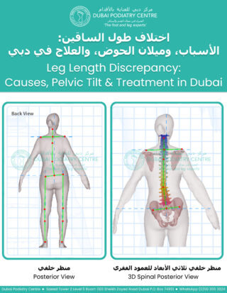
Dubai Podiatry Centre graphic titled “Leg Length Discrepancy: Causes, Pelvic Tilt & Treatment in Dubai,” with a rear-view body alignment diagram and a 3D rear view of the spine and pelvis

This raises the foot more uniformly rather than concentrating the adjustment beneath the heel.

The correct choice depends on the cause and size of the discrepancy, the patient’s foot mechanics, footwear and symptoms.

For this reason, we recommend a thorough biomechanical assessment before deciding whether a heel raise, full-length lift or corrective orthotic is required.

Could Correcting a Collapsed Arch Improve an Apparent Leg Length Difference?

In certain patients, the apparent difference in leg length is partly related to excessive pronation or collapse of the medial longitudinal arch.

When this occurs, supporting and controlling the foot may improve the way the lower limb functions.

A corrective orthotic may help improve foot positioning and reduce some of the compensatory movements occurring further up the body.

In selected cases, this can reduce the functional component of an apparent leg length discrepancy.

However, arch correction does not lengthen a physically shorter femur or tibia.

This distinction is essential when choosing the most appropriate treatment.

Assessing Leg Length Discrepancy With 3D Body Scanning

At Dubai Podiatry Centre, we can incorporate 3D body scanning technology into a comprehensive biomechanical assessment.

Our infrared-based scanner can help us evaluate visible postural alignment and asymmetries, including:

  • Shoulder height differences
  • Pelvic positioning
  • Apparent lower-limb asymmetry
  • Foot posture and alignment
  • Overall standing posture

Infrared scanning does not use ionising radiation, making it a useful non-invasive assessment tool for adults and children.

However, it is important to understand that a surface body scanner cannot directly measure the length of the femur or tibia, diagnose hip dysplasia or replace medical imaging when an accurate structural measurement is required.

Clinical examination, gait analysis and, when indicated, appropriate imaging are used together to establish the underlying cause.

Leg Length Discrepancy in Children

Children may develop apparent or structural leg length differences for several reasons.

These may include differences in bone growth, foot posture or conditions affecting the hip.

Developmental dysplasia of the hip is one condition that must be considered when assessing certain children with asymmetry.

Although newborn screening is routinely performed, some hip conditions may still require further investigation later in childhood.

A child with persistent limping, significant asymmetry, hip discomfort or a suspected structural discrepancy should be referred for appropriate paediatric or orthopaedic assessment.

Progressive Corrective Orthotics

At Dubai Podiatry Centre, we use a progressive approach to corrective orthotic treatment where clinically appropriate.

Rather than introducing a substantial biomechanical correction immediately, adjustments can be made in carefully planned stages.

This allows the feet and lower limbs time to adapt to changes in support and alignment.

Our treatment approach may include:

1. A comprehensive assessment of foot and ankle biomechanics.
2. Evaluation of apparent and structural leg length differences.
3. Assessment of gait and pelvic alignment.
4. Custom-made orthotics where indicated.
5. Progressive adjustments based on the patient’s response.
6. Follow-up assessments to monitor comfort, function and alignment.

Not every patient requires orthotics, and not every leg length discrepancy needs correction.

The objective is to provide an appropriate level of support and correction for each individual.

When Should You Have Your Leg Length Checked?

You may benefit from a biomechanical assessment if you experience persistent symptoms such as:

  • One-sided lower back or hip discomfort
  • A feeling that one leg is longer than the other
  • Uneven wear on your shoes
  • One foot arch appearing significantly flatter than the other
  • Recurrent lower-limb discomfort during walking or sport
  • Noticeable asymmetry in standing posture
  • Persistent pain, progressive weakness, numbness or significant walking difficulties should also be assessed by an appropriate medical professional.

The Importance of Looking at the Whole Body

A leg length discrepancy is not always simply a question of one leg being too short.

Sometimes the difference is structural. Sometimes it is functional. Occasionally, both factors are present.

Understanding the relationship between the foot arches, ankles, knees, pelvis and spine is an important part of identifying the underlying problem.

At Dubai Podiatry Centre, our approach is to assess the biomechanics of the lower limbs and develop an individualised treatment plan where correction is clinically indicated.

If you are concerned about uneven leg lengths, collapsed arches or the way your feet may be affecting your posture, contact Dubai Podiatry Centre to arrange a comprehensive biomechanical assessment.

Website: www.dubaipodiatry.com

For more information or to book an appointment please call our clinic +971 4 3435390 or WhatsApp +971 50 3553024


When we think about the arches of our feet, most people immediately picture the large arch running along the inside of the foot.

But there is another, much smaller arch at the front of the foot that we pay very close attention to during a biomechanical assessment at Dubai Podiatry Centre.

It is called the transverse arch.


What is the transverse arch?

The front part of your foot is called the forefoot. It includes the toes and the metatarsal bones, which are the long bones immediately behind your toes.

Across the forefoot, just behind the toes where they connect with the foot, is the transverse arch.

Rather than running from the heel towards the toes like the more familiar medial arch, the transverse arch runs across the foot from side to side.

It is a relatively small arch, but biomechanically it can be extremely important.


What happens when the transverse arch starts to drop?

Over time, the forefoot can begin to spread and the transverse arch can become lowered.

We sometimes describe this to patients as the metatarsal bones beginning to “avalanche” forwards and downwards.

Although this change is occurring in a relatively small area at the front of the foot, it can influence the way the rest of the foot functions.

The metatarsal bones do not work independently from the remainder of the foot. They form part of the interconnected structure of the foot and contribute to its stability and function during standing and walking.

As the forefoot begins to change position, we may also see changes occurring through the larger medial arch, the midfoot and eventually the mechanics around the ankle.

This is why we never look at the transverse arch in isolation.


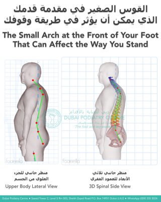
3D postural assessment showing how a collapsed forefoot transverse arch may affect standing posture and spinal alignment


Are you beginning to look hunched forwards?

One of the observations that can prompt us to investigate the forefoot is a change in a person’s stature.

Sometimes a patient, or their family, will say that they are beginning to “look hunchbacked” or that they appear to be leaning forwards.

The head may be sitting further forwards, the shoulders may appear more rounded and the person can develop an increasingly stooped appearance.

There are, of course, many different reasons why somebody may develop a forward head position or stooped posture. It should never automatically be assumed that it is coming from the feet.

However, it is one of the physical signs that makes us look more carefully at the person’s overall biomechanics.

We look at the person from the side and assess their stature. We look at the position of the head and shoulders. We observe how they stand and walk.

Then we investigate what is happening underneath them.

In particular, we examine the sole of the foot and the forefoot to see what has happened to the transverse arch.

If that small arch has dropped and the metatarsal bones have moved forwards and downwards, it may be contributing to a chain of biomechanical changes occurring further up the body.

Your feet form the foundation of your posture


The feet provide the foundation for the body when we stand and walk.

Changes in that foundation can sometimes contribute to compensations further up the biomechanical chain.

This is why a person’s posture can give us clues about what we should investigate in their feet.

A forward head, rounded shoulders, a stooped stance or a tendency to shuffle when walking does not diagnose a foot problem. However, these observations can be a reason to include the feet in the assessment rather than concentrating only on the upper body.

Sometimes the clue is visible at the shoulders and head, while part of the biomechanical problem may be much further down, at the sole of the foot.


Why we look for problems early

One of the reasons we pay such close attention to the transverse arch is that it can give us an opportunity to identify biomechanical changes relatively early.

If the transverse arch has started to lower but the remainder of the foot is still relatively well controlled, treatment can be much simpler.

Where appropriate, a custom corrective orthotic can be designed to support and lift the transverse arch and improve the mechanical stability of the forefoot.

The objective is not simply to put something soft underneath a painful area. It is to address the position and function of the forefoot as part of the mechanics of the entire foot.


What if the whole foot has already started to collapse?

More advanced biomechanical changes can also be managed. It simply requires a different approach.

If changes have progressed through the forefoot, midfoot and ankle, attempting to correct everything at once may not be appropriate or comfortable.

Instead, orthotic therapy may need to be carried out step by step and stage by stage.

We may initially need to address the forefoot and transverse arch. As the foot adapts, we can reassess the midfoot, medial arch, rearfoot, heel and ankle mechanics.

This process naturally takes longer than treating an early biomechanical change.

A small arch can give us a big clue

The transverse arch is a relatively small structure, but it can tell us a great deal about what is happening throughout the foot.

This is why we don’t assess feet simply by asking, “Where does it hurt?”

We look at the toes, forefoot, midfoot, heel and ankle. We look at how you stand. We look at the position of your shoulders and head. We watch how you walk.

If somebody tells you that you are beginning to look more hunched over, or you notice that your head is increasingly sitting forwards and your shoulders are becoming rounded, there can be many possible explanations.

But don’t forget to look down.

A biomechanical assessment of the feet, and particularly that small transverse arch underneath the forefoot, may provide another important piece of the puzzle.

The earlier we identify a change in foot biomechanics, the easier it may be to manage before it progresses further.


Dubai Podiatry Centre
Biomechanical Foot Assessment and Custom Orthotic Therapy


For more information or to book an appointment please call our clinic +971 4 3435390 or WhatsApp +971 50 3553024



Have you ever wondered how custom-made corrective orthotics can transform the way you stand and walk? Watch our before-and-after video to see the remarkable improvement in foot alignment after wearing custom orthotics designed specifically to correct overpronation and flat feet.


When the feet lean inward excessively, they can cause poor body alignment, placing extra pressure on the ankles, knees, hips, and lower back. This imbalance often contributes to heel pain, plantar fasciitis, arch pain, ankle instability, knee pain, corns, calluses, and foot fatigue. Without proper support, these problems can worsen over time and affect everyday mobility.

At Dubai Podiatry Centre, our experienced podiatrists perform a comprehensive biomechanical assessment, gait analysis, and foot impression to accurately diagnose the cause of your foot pain. Using these findings, we design custom-made corrective orthotics that help restore proper foot posture, improve balance, reduce excessive pressure, and support natural walking mechanics.

This before-and-after transformation highlights how corrective orthotics can improve foot positioning, enhance stability, and promote healthier movement. By supporting the feet in a more neutral position, custom orthotics help reduce strain throughout the body and improve overall comfort during walking, standing, and daily activities.

If you suffer from flat feet, overpronation, heel pain, plantar fasciitis, foot pain, ankle pain, knee pain, arch pain, or recurring corns and calluses, a professional biomechanical assessment may be the first step towards lasting relief.

Book an appointment with Dubai Podiatry Centre to discover how custom corrective orthotics can improve your foot alignment, relieve pain, and help you move with greater confidence.

For more information or to book an appointment please call our clinic +971 4 3435390 or WhatsApp +971 50 3553024
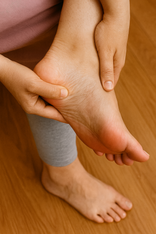


Many people think of foot problems as simply aches, pains, or cosmetic concerns, but your feet can provide valuable insight into your overall health and longevity.

Your feet are often one of the first places where signs of health problems become noticeable. Poor circulation, nerve damage, inflammatory conditions, and biomechanical abnormalities can all affect the feet long before more serious symptoms develop elsewhere in the body.

For example, elevated uric acid levels can lead to gout, which commonly affects the big toe joint and can cause sudden episodes of severe pain and inflammation. Diabetes and elevated blood glucose levels can damage nerves and blood vessels, leading to numbness, tingling, reduced sensation, and poor wound healing in the feet. Circulatory problems, often associated with smoking, cardiovascular disease, or narrowed blood vessels, can also be detected during a foot assessment.

The structural health of your feet is equally important. When the feet and ankles are not properly aligned, the body must compensate during walking. One common example is excessive inward rolling of the ankles, which can cause the feet to point outward during gait. This altered walking pattern reduces efficiency and can affect the activation of important muscle groups, including the calf muscles.

The calf muscles are often referred to as the body’s “second heart” because they help pump blood from the legs back towards the heart. Healthy foot and ankle function promotes efficient walking, better calf muscle activity, improved circulation, and greater overall mobility.

Research consistently shows that maintaining mobility, balance, strength, and regular physical activity are important factors associated with healthy ageing and longevity. Since every step begins with your feet, keeping them healthy and functioning correctly is an essential part of maintaining an active lifestyle.

If you have foot pain, ankle instability, poor balance, numbness, circulation concerns, or simply want to understand how your feet may be affecting the rest of your body, consider a comprehensive podiatry assessment. A detailed biomechanical, circulatory, and neurological examination can provide valuable information about your foot health and overall wellbeing.

Your feet carry you through life, looking after them may help you stay active, independent, and healthier for longer.


For more information or to book an appointment please call our clinic +971 4 3435390 or WhatsApp +971 50 3553024


Why Plantar Fasciitis Isn’t Really About a “Tight” Fascia

By Michelle Champlin, Chief Podiatrist – Dubai Podiatry Centre

Plantar fasciitis is one of the most common and painful foot conditions we see in clinic. It causes sharp, stabbing heel pain—especially when getting out of bed in the morning or after sitting for a while. But the name plantar fasciitis can actually be misleading.


Understanding the Plantar Fascia

The plantar fascia is a thick, collagen-like band that runs from the heel bone to the toes. It acts like a suspension bridge to keep the foot’s arches intact and to stabilise every step you take.

Because the plantar fascia is made of densely woven collagen fibres, it’s extremely strong but not very stretchy. Many articles suggest “stretching” the fascia, but in truth, it doesn’t tighten—it becomes pulled taut when the foot’s structure begins to collapse.

Why It Happens

When you have healthy arches, the plantar fascia works quietly in the background to keep your foot stable. But when one or more of your foot’s three main arches—

  • the medial arch (inside edge of the foot),
  • the lateral arch (outer edge), or
  • the transverse arch (across the ball of the foot)—
  • start to flatten or your ankle leans inward or outward, the fascia is forced to work overtime.

That constant tension at its attachment into the heel bone causes tiny micro-tears in the outer casing of the bone, leading to inflammation and pain.

Why the Pain Comes and Goes

When you sit or sleep, inflammation builds up in the casing around the heel bone. The moment you stand up, pressure on that area causes intense pain. After walking a few minutes, your calf muscles begin to pump blood through the foot, draining the inflammation and easing the discomfort—until you sit again.

That’s why many people describe their first steps out of bed as agonising, but feel somewhat better once they “get moving.”


Who Gets Plantar Fasciitis—And Who Doesn’t

Interestingly, people with genuinely flat feet rarely get plantar fasciitis, because their fascia never needs to engage—it’s already at rest. The condition typically affects those whose arches are collapsing under load, not those whose arches are already fully flat.

The Real Solution: Structural Correction

Treating the heel pain directly—ice, massage, or anti-inflammatory creams—only addresses the symptom. The long-term solution is to correct the structure of the foot so that the fascia no longer has to overwork.

This might involve:

  • Custom orthotics to lift the medial and transverse arches
  • Supporting the ankle to stop it leaning in or out
  • Strengthening the calf and intrinsic foot muscles


Once the arches are properly supported, the fascia relaxes and the inflammation at the heel naturally subsides.


Healing Time: 12–14 Weeks

True healing takes time. The recovery process for plantar fasciitis typically spans 12 to 14 weeks.

You can imagine the heel bone where the plantar fascia attaches as a large window that has had a stone thrown at it, causing tiny micro-fractures across the glass.

  • During the first six weeks, the outer edges of the “window” begin to self-heal.
  • Over the next six weeks, the centre, or the “bull’s-eye” where the impact was greatest, gradually repairs.
  • The final two weeks allow for complete healing and realignment.

This is why genuine corrective orthotic therapy, which lifts and supports all three arches of the foot and stabilises the ankle, requires this full 12–14 week period—to give the plantar fascia the rest it needs for complete recovery.

A Helpful Analogy

Imagine the plantar fascia like a strong sticker on a wall. If you start to peel it off, some paint and plaster may come away, but the concrete beneath is still intact. That’s similar to what happens when the fascia pulls on the heel bone—micro-damage occurs, but the structure can heal once tension is reduced.

Another way to picture it: think of the heel bone as a ceiling with a hidden water bubble. When you sleep, fluid builds up in that bubble, and when you stand, it’s suddenly compressed—painful until movement drains it away.

In Summary

Plantar fasciitis isn’t simply inflammation of a “tight” band. It’s a structural response to stress in the foot’s architecture. Correct the mechanics, and the pain goes away naturally.

If you suffer from heel pain, especially first thing in the morning, book a biomechanical assessment with our podiatrists. By identifying which arch is collapsing, we can design a treatment that targets the cause—not just the symptom.


#PlantarFasciitis #HeelPain #HeelPainRelief #DubaiPodiatryCentre #FootPain #FlatFeet #ArchSupport #OrthoticsDubai #Podiatry #FootCare #Biomechanics #HealthyFeet #Footerella #PodiatristDubai #DubaiHealth


For more information or to book an appointment please call our clinic +971 4 3435390 or WhatsApp +971 50 3553024


Copyright Dubai Podiatry 2026. All rights reserved.

WhatsApp us