

fbpx

Please wait...


BLOG

No more posts

One of the most common questions we hear at Dubai Podiatry Centre is: “Why won’t my nail fungus clear, even though I’ve tried home remedies?”

The answer is that not all fungal nail infections are the same.

Different types of nail fungus affect different parts of the toenail, and each type requires a different treatment approach. This is why a treatment that works for one person may have little or no effect for another.


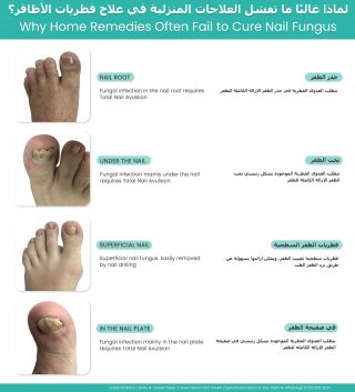
Why home remedies often fail to cure nail fungus showing fungal infections in the nail root, under the nail, superficial nail fungus, and nail plate with appropriate treatment options

Superficial White Nail Fungus

One of the easiest types of fungal infection to treat is superficial white fungal infection.

This commonly develops in warm, humid conditions inside closed footwear and under nail polish. The fungus affects only the surface of the nail and often appears as white, chalky or powdery patches on the top of the toenail.

Because the infection is only on the surface, the affected layer can often be gently reduced with professional nail filing or careful buffing, allowing a topical antifungal nail lacquer to reach the remaining fungus more effectively.

As the nail gradually grows out, the healthy nail replaces the infected portion.


Fungus Growing Beneath the Nail

A more difficult type of fungal infection develops underneath the nail plate.

This is commonly associated with a widespread fungal skin infection affecting the sole of the foot, often referred to as moccasin-type athlete’s foot. The fungus can spread beneath the toenail, where it becomes protected by the thick nail plate.

When the fungus is trapped underneath the nail, simply painting antifungal medication onto the surface often has limited success because the medication cannot adequately reach the infection.


Depending on the severity of the infection, treatment may involve oral antifungal medication, careful reduction of the thickened nail, or, in selected cases, nail avulsion of the toenail to allow direct treatment of the underlying nail bed as the new nail grows.

Following nail removal, the smaller toenails generally regrow in approximately three months, while the big toenail may take anywhere from six months to one year to fully regrow.


Fungus That Reaches the Nail Root

In some patients, the fungal infection extends into the nail root (matrix), where the nail is produced.

When this occurs, oral antifungal medication is often recommended because the medication reaches the growing nail through the bloodstream, allowing the new nail to grow free of infection.

If there is also extensive fungal material trapped beneath a thick or damaged nail plate, reducing or removing the affected nail may be recommended to improve access for topical treatment and encourage healthy nail regrowth.


When Oral Medication Is Not Suitable

Before prescribing oral antifungal medication, liver function blood tests are often performed to ensure the medication can be taken safely.

Occasionally, these tests identify previously undiagnosed liver abnormalities. In these situations, even patients who feel completely healthy may not be suitable candidates for oral antifungal medication.

For these patients, treatment may focus on physical methods, including removal of the infected toenail where appropriate, combined with topical antifungal medication applied directly to the exposed nail bed after healing. This allows the new nail to grow in an environment with a much lower fungal burden.


Why Professional Assessment Matters

Many people spend months or even years trying home remedies without success, simply because they are treating a different type of fungal infection.

Successful treatment begins with identifying exactly where the fungus is located, whether it is on the surface of the nail, beneath the nail plate, or involving the nail root.

Once the type and stage of the infection have been identified, the most appropriate treatment plan can be chosen, giving the best opportunity for healthy, fungus-free nail growth.


For more information or to book an appointment please call our clinic +971 4 3435390 or WhatsApp +971 50 3553024
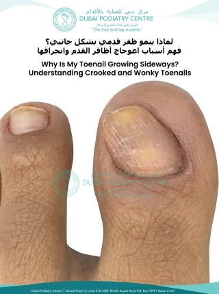


One of the most common questions patients ask at Dubai Podiatry Centre is:

“Am I too old to treat my flat feet?”

The simple answer is no.

Many people believe that flat feet can only be treated during childhood and that once you become an adult, nothing more can be done. Fortunately, this is one of the biggest misconceptions we hear in clinic.


Not Everyone Diagnosed with Flat Feet Actually Has Flat Feet

There are several different types of flat feet, but the most common condition we see is not a true flat foot at all.

In many cases, the ankle leans inwards, causing the arch to collapse under body weight. The arch then appears flat, even though the foot itself still has the ability to regain its natural shape once the ankle is correctly aligned.

Rather than thinking of the foot as permanently flat, it is often more accurate to think of it as an arch that has collapsed because the ankle is no longer supporting it correctly.


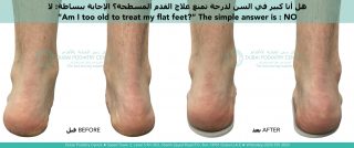
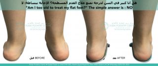
Adult flat feet before and after custom orthotics showing improved heel alignment and foot posture

Child with flat feet before and after podiatry treatment using custom orthotics showing corrected heel alignment


Adult Treatment Can Actually Be Faster

Many people are surprised when we explain that correcting this type of flat foot can actually progress faster in adults than in children.

Children have high levels of human growth hormone, which keeps their ligaments soft, flexible and easy to manipulate. This makes repositioning the bones relatively straightforward and usually very comfortable.

However, because the ligaments are constantly growing and remodelling, they often take longer to adapt and stabilise in their new position. For this reason, children commonly progress through each stage of treatment over approximately three to six months.

Adults are different.

Adult ligaments are firmer and less elastic, so there can be slightly more resistance when repositioning the foot. For this reason, we usually make smaller adjustments at each stage.

Once the bones have been repositioned, however, the adult foot often adapts surprisingly quickly. Instead of waiting three to six months between stages, many adults can progress every one to three months, allowing treatment to advance more rapidly.

Similar to Orthodontic Braces

A useful comparison is orthodontic braces for the teeth.

Most people understand that braces are not just for children. Adults successfully wear braces every day to gradually reposition their teeth into better alignment.

Corrective orthotics work on a similar principle.

Instead of moving teeth, they gently reposition the bones of the feet and ankles in carefully planned stages. As the alignment improves, the arch is supported more effectively and, in suitable cases, the foot can gradually regain a healthier arch shape.

Each new stage builds upon the previous one, allowing gradual correction rather than trying to make one large adjustment all at once.


It’s Never Too Late to Have an Assessment

If you have been told you have flat feet, it does not necessarily mean your feet are permanently flat.

The first step is determining why your arch has collapsed. For many patients, the underlying issue is ankle alignment rather than the shape of the foot itself.

With the correct diagnosis and a staged treatment plan, adults can often achieve significant improvements in foot alignment, comfort and function.

So if you’ve been wondering whether you’ve left it too late to treat your flat feet, the answer may be much more encouraging than you expected.

At Dubai Podiatry Centre, we regularly assess both children and adults and develop personalised treatment plans based on the specific cause of each patient’s foot alignment, because no two cases of flat feet are exactly the same.


For more information or to book an appointment please call our clinic +971 4 3435390 or WhatsApp +971 50 3553024
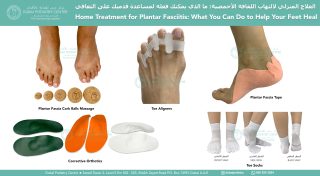


Plantar fasciitis is one of the most common causes of heel pain. People typically notice it when they take their first steps out of bed in the morning or when they stand after sitting for a prolonged period. The pain often eases after walking for a few minutes, only to return again after periods of rest.


What is the plantar fascia?

The plantar fascia is a strong band of connective tissue that runs from the heel bone (calcaneus) to the base of the toes. Its primary function is to help support the arch of the foot.

When you walk, run or land from a jump, the plantar fascia becomes tensioned, helping the arch maintain its shape and absorb forces. Without this support, the bones of the midfoot would be subjected to much greater mechanical stress.

A healthy plantar fascia is not designed to remain under constant tension. Instead, it should be relatively relaxed at rest and become taut only when required during movement and weight-bearing activities.


Why does plantar fasciitis develop?

The term plantar fasciitis literally means inflammation associated with the plantar fascia. Although the pain is felt in the heel, the discomfort is usually concentrated where the plantar fascia attaches to the heel bone.

Rather than viewing plantar fasciitis as simply an isolated heel problem, it is often more helpful to consider why the plantar fascia has become overloaded in the first place.

In many people, the ligaments and muscles of the foot provide enough stability to support the arch without excessive strain on the plantar fascia. However, if the alignment of the foot changes, the plantar fascia may remain under tension for much longer than it was designed to.

One of the most common contributing factors is excessive pronation of the foot and ankle. As the foot rolls inward, the plantar fascia may become progressively tensioned throughout standing and walking instead of only during the phases when it is normally required.

An easy way to understand the problem

Imagine holding a single strand of long hair with a heavy weight attached to the end.

The hair itself is not painful.

The pain would occur where the hair is anchored into the scalp because that attachment point is constantly being pulled.

Once the weight is removed, the strain at the attachment point disappears.

The plantar fascia behaves in a similar way. The discomfort usually develops where the fascia attaches into the heel bone rather than within the fascia itself. If excessive tension continues day after day, this attachment site can become irritated and painful.


Why is the pain worse in the morning?

Many people notice that the first few steps in the morning are the most painful.

After sleeping or sitting for a prolonged period, blood flow and tissue fluid movement slow down. When the foot is suddenly loaded, the already irritated attachment of the plantar fascia is placed under tension again, producing the characteristic sharp heel pain.

As you begin walking, the calf muscles act as a pump, improving circulation through the lower leg and foot. This often reduces discomfort temporarily, which explains why many people feel better after several minutes of walking.

Treating the cause—not just the symptoms

While heel cushions, supportive insoles and stretching programmes may provide symptom relief for some individuals, long-term improvement depends on identifying and correcting the mechanical cause of the overload.

If excessive pronation or another alignment abnormality is placing unnecessary tension on the plantar fascia, simply adding cushioning beneath the heel may not adequately address the underlying problem.

A corrective orthotic is designed to improve foot and ankle alignment rather than simply support the arch. Because every person walks differently, the correction required for the left foot is often different from that required for the right foot. The amount of correction should therefore be individually prescribed.

As the abnormal strain on the plantar fascia is reduced over time, the attachment at the heel is given the opportunity to recover, allowing symptoms to gradually settle while also addressing the underlying biomechanical problem that contributed to the condition.

The key message

Plantar fasciitis should not simply be viewed as inflammation of the heel. In many cases, it is the result of an underlying mechanical imbalance that places excessive and prolonged tension on the plantar fascia where it attaches to the heel bone.

Successful long-term treatment focuses on identifying the biomechanical cause, correcting the alignment of the foot and ankle, and reducing the abnormal forces responsible for the condition. By addressing the underlying mechanics rather than simply cushioning the symptoms, many patients can achieve lasting relief and reduce the likelihood of recurrence.


For more information or to book an appointment please call our clinic +971 4 3435390 or WhatsApp +971 50 3553024
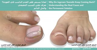


Many people think of foot problems as simply aches, pains, or cosmetic concerns, but your feet can provide valuable insight into your overall health and longevity.

Your feet are often one of the first places where signs of health problems become noticeable. Poor circulation, nerve damage, inflammatory conditions, and biomechanical abnormalities can all affect the feet long before more serious symptoms develop elsewhere in the body.

For example, elevated uric acid levels can lead to gout, which commonly affects the big toe joint and can cause sudden episodes of severe pain and inflammation. Diabetes and elevated blood glucose levels can damage nerves and blood vessels, leading to numbness, tingling, reduced sensation, and poor wound healing in the feet. Circulatory problems, often associated with smoking, cardiovascular disease, or narrowed blood vessels, can also be detected during a foot assessment.

The structural health of your feet is equally important. When the feet and ankles are not properly aligned, the body must compensate during walking. One common example is excessive inward rolling of the ankles, which can cause the feet to point outward during gait. This altered walking pattern reduces efficiency and can affect the activation of important muscle groups, including the calf muscles.

The calf muscles are often referred to as the body’s “second heart” because they help pump blood from the legs back towards the heart. Healthy foot and ankle function promotes efficient walking, better calf muscle activity, improved circulation, and greater overall mobility.

Research consistently shows that maintaining mobility, balance, strength, and regular physical activity are important factors associated with healthy ageing and longevity. Since every step begins with your feet, keeping them healthy and functioning correctly is an essential part of maintaining an active lifestyle.

If you have foot pain, ankle instability, poor balance, numbness, circulation concerns, or simply want to understand how your feet may be affecting the rest of your body, consider a comprehensive podiatry assessment. A detailed biomechanical, circulatory, and neurological examination can provide valuable information about your foot health and overall wellbeing.

Your feet carry you through life, looking after them may help you stay active, independent, and healthier for longer.


For more information or to book an appointment please call our clinic +971 4 3435390 or WhatsApp +971 50 3553024
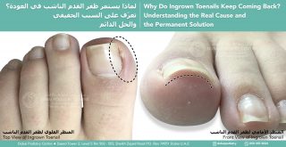


A patient attended our clinic today complaining of severe pain underneath the outside of his left foot. The pain had become so intense that he was limping and described the sensation as walking on a sharp piece of glass.

On examination, we found a hard corn underneath the fifth metatarsal head on the outside border of his left foot. Many people assume that a corn is the problem, but in reality a corn is often the body’s attempt to protect itself from excessive pressure.

We removed the corn painlessly, and the patient experienced immediate relief. However, we explained that unless we addressed the underlying cause, the corn would simply return.

To investigate further, we performed a 3D body scan and biomechanical assessment.

The results were fascinating.

The patient had a significant imbalance throughout his body. His left leg was functionally longer than his right. His pelvis was tilted, with the right side anteriorly tilted and the left side posteriorly tilted. He was carrying considerably more body weight through the left side of his body, creating excessive pressure beneath the outside of the left foot.

His medical history helped explain the pattern. He had previously undergone two lower back surgeries on the left side, suffered left hip problems, torn ACL injuries involving the left knee, and undergone left Achilles tendon surgery.

Over many years, his body had adapted to these injuries.

The scan showed that his pelvis had shifted towards the left side, causing increased loading through the left leg and foot. At the same time, his right foot was internally rotated and functioning almost like a golfer at the top of a backswing position. This created high tensile stress through the right Achilles tendon, the right knee, and the lower back while simultaneously increasing compression forces through the left side of the body.

The painful corn underneath the left foot was simply the area where the body was absorbing the greatest amount of pressure.

The development of a corn follows a predictable sequence.

First, the body produces a layer of hard skin to protect itself. This creates a broad plaque of callus, similar to a pancake of thicker skin.

If the pressure continues, the body creates a concentrated plug of dense keratin at the centre of the pressure point. Keratin is the same material found in toenails. This hard central core forms what we recognise as a hard corn.

As pressure continues to increase, inflammation develops beneath the area. Patients often describe burning, stinging, or sharp sensations. This was the stage our patient had reached.

If left untreated, the next stage can be the formation of a soft corn. The tissues become saturated with inflammatory fluid and appear white, rubbery, and extremely painful.

The final stage is tissue breakdown and ulceration.

This progression is particularly important in patients with diabetes, neuropathy, or vascular disease. These patients may not feel the early warning signs of pressure and pain. As a result, the protective stages can progress unnoticed until an ulcer develops.

Many patients believe ulcers appear suddenly and without warning. In reality, the body often gives multiple warning signals beforehand, but if sensation is reduced, those signals may not be recognised.

For this patient, the solution was not repeated corn removal.

The solution was correcting the forces causing the corn.

We prescribed custom corrective orthotics designed specifically for his individual biomechanics. The right orthotic was intentionally very different from the left because each side of the body required a different correction.

The aim was to improve pelvic balance, reduce the functional leg length discrepancy, redistribute pressure more evenly between both feet, and decrease excessive loading through the left foot.

We performed further scans with the orthotics in place and saw immediate improvements in alignment and pressure distribution.

The body can be compared to a wobbly table. If one table leg is shorter than the others, the tabletop becomes uneven. Placing a carefully measured support beneath the shorter leg restores balance.

Corrective orthotics work in a similar way. While we cannot make one leg physically grow longer, we can improve balance and alignment so that the body functions more efficiently and with less stress.

The patient will return in three months for reassessment. As the body adapts to improved alignment, muscle tension, posture, and weight distribution can all change. Repeat scanning will allow us to monitor these changes and determine whether further orthotic adjustments are required.

The important lesson from this case is that a corn is rarely just a skin problem.

A corn is often a warning sign that excessive pressure is travelling through a specific part of the foot. Removing the corn may provide temporary relief, but identifying and correcting the underlying mechanical cause is what provides a long-term solution.


For more information or to book an appointment please call our clinic +971 4 3435390 or WhatsApp +971 50 3553024


One of the most common reasons people delay seeking treatment for toenail problems is simple embarrassment.

Many patients tell us they have been hiding their feet for years. They avoid sandals, swimming pools, pedicures, and sometimes even showing their feet to family members. Some have been covering their toenails with nail polish or avoiding treatment altogether because they feel self-conscious about how their nails look.

The truth is that podiatrists see toenail problems every single day.

Whether your toenails are thickened, discoloured, fungal, damaged, ingrown, lifted, crumbly, misshapen, or have been neglected for years, there is no judgment. Our goal is to help you restore the health of your feet and regain confidence in your appearance.

Many toenail conditions are much more common than people realise. Fungal infections, bacterial infections, trauma from sports, injuries, and poorly fitting footwear can all affect the appearance and health of the toenails. Sometimes a single accident or even one particular pair of shoes can start a problem that gradually worsens over time.

The good news is that treatment options are available.

Depending on the condition, treatment may involve:

  • Professional nail care and reduction
  • Antifungal treatments
  • Oral medication when appropriate
  • Treatment of bacterial infections
  • Footwear recommendations
  • Toe socks to improve toe separation and reduce fungal recurrence (link for toe socks)
  • Temporary or permanent nail procedures when necessary


Sometimes the treatment plan may sound dramatic at first. For example, a severely damaged or chronically infected toenail may occasionally need to be removed to allow a healthy nail to regrow. However, this is often the quickest route back to a healthy, comfortable, and attractive nail.

Before treatment of fungal toenail infection showing a damaged big toenail with yellow discoloration, nail thickening, crumbling debris, and signs of onychomycosis.

Severe fungal toenail infection before treatment showing thickened, yellow-brown, brittle nail with extensive nail damage and discoloration.

Fortunately, toenails are remarkably good at regenerating. Smaller toenails can often regrow within approximately three months following removal, while larger toenails may take six months to a year to fully grow back, depending on the individual and the size of the nail. In the context of a lifetime of healthy feet, this is a relatively short period of time.


At your consultation, your podiatrist will assess the cause of the problem and develop a personalised treatment plan designed to restore the health and appearance of your toenails. Every patient is different, and treatment should be tailored to the individual.

Most importantly, everything discussed during your appointment is completely confidential. We understand that many people feel anxious or embarrassed about their feet, but there is no need to suffer in silence.


Our job is to help you get your feet back to how you remember them being—or perhaps even better than before.


So if you have been hiding your feet, postponing treatment, or feeling embarrassed about your toenails, don’t wait any longer. Make an appointment with a podiatrist and take the first step towards healthier feet and greater confidence.


For more information or to book an appointment please call our clinic +971 4 3435390 or WhatsApp +971 50 3553024


Many people are surprised to learn that fungal toenail infections often begin with a fungal skin infection between the toes, commonly known as athlete’s foot (tinea pedis).


The fungi responsible for athlete’s foot are commonly found in our environment and can also exist naturally on the skin without causing problems. However, when conditions become warm, dark, and moist, the fungal organisms can multiply rapidly. This is especially common between the fourth and fifth toes, where the skin surfaces are often pressed tightly together inside shoes.


Multiple toes affected by fungal toenail infection (onychomycosis) showing yellow, thickened, and discolored toenails. Clinical image used to discuss whether toe socks can help prevent fungal toenail infections and improve foot hygieneWhen the toes are squeezed together for long periods, moisture becomes trapped and air circulation is reduced. This creates the perfect environment for fungal spores to grow and spread. Over time, the infection may remain confined to the skin between the toes, or it may spread into the neighbouring toenails, particularly the fourth and fifth toenails, resulting in fungal nail infection (onychomycosis).


One simple but highly effective preventative measure is the use of cotton toe socks.


Unlike conventional socks, toe socks individually separate each toe with a layer of fabric. This creates a physical barrier between the toes, helping to reduce skin-to-skin contact and minimise the warm, moist conditions that fungi thrive in.

The benefits of toe socks include:

  • Improved air circulation between the toes
  • Reduced moisture accumulation
  • Less skin friction and irritation
  • A physical barrier that helps limit the spread of fungal spores
  • Reduced recurrence of athlete’s foot
  • Lower risk of fungal infection spreading into the toenails


Cotton toe socks are particularly beneficial for individuals who suffer from recurring athlete’s foot, fungal toenail infections, excessive sweating, or who spend long periods wearing enclosed footwear.


While toe socks are not a treatment for an established fungal infection, they can be an excellent part of a long-term prevention strategy. Combined with good foot hygiene, proper shoe disinfection, and prompt treatment of any athlete’s foot infection, toe socks can help create a healthier environment for the feet and reduce the likelihood of future fungal problems.


At Dubai Podiatry Centre, we frequently recommend toe socks as part of a comprehensive foot care programme for patients prone to athlete’s foot and fungal toenail infections. Sometimes the simplest changes can make a significant difference in maintaining healthy feet.


Link for : Toe Socks


For more information or to book an appointment please call our clinic +971 4 3435390 or WhatsApp +971 50 3553024


Dermojet device used for plantar wart treatment, showing a needle-free high-pressure injector designed to deliver medication into deep verrucas on the sole of the foot.At Dubai Podiatry Centre, our highest success rate for plantar warts comes from our Dermojet treatment.

Dermojet uses a high-velocity stream of liquid delivered through a mechanical device. When the pressure is released, a microjet of liquid is ejected from the apparatus and penetrates through the skin to reach the deeper part of the plantar wart, allowing the treatment to be delivered more efficiently.

Warts elsewhere on the body usually grow outward. However, warts on the sole of the foot grow inward because of pressure from standing and walking. This means that treatments such as liquid nitrogen, salicylic acid, and laser, which may work well on external warts on other parts of the body, can have very different outcomes when used on the sole of the foot.

في مركز دبي لطبّ القدم، نحقق أعلى نسبة نجاح في علاج الثآليل الأخمصية (ثآليل باطن القدم) باستخدام علاج الديرموجيت (Dermojet).

يعتمد الديرموجيت على إطلاق تيار عالي السرعة من السائل عبر جهاز ميكانيكي. وعند تحرير الضغط، يُقذف تيار دقيق من السائل من الجهاز ليخترق الجلد ويصل إلى الجزء الأعمق من الثؤلول الأخمصي، مما يسمح بإيصال العلاج بكفاءة أكبر وفعالية أعلى.

عادةً ما تنمو الثآليل في مناطق أخرى من الجسم إلى الخارج. أما الثآليل الموجودة في باطن القدم فتنمو إلى الداخل بسبب الضغط الناتج عن الوقوف والمشي. وهذا يعني أن العلاجات مثل النيتروجين السائل، وحمض الساليسيليك، والليزر، والتي قد تكون فعّالة في علاج الثآليل الخارجية في أجزاء أخرى من الجسم، قد تعطي نتائج مختلفة تمامًا عند استخدامها لعلاج الثآليل الموجودة في باطن القدم.

Before and after plantar wart treatment using Dermojet showing wart resolution on sole of foot


A wart on the top of the hand, for example, is more exposed and easier to reach with acid or liquid nitrogen. A plantar wart, commonly called a verruca, is inverted into the sole of the foot and is therefore less accessible.

For this reason, Dermojet is mainly reserved for plantar warts and verrucas on the sole of the foot. It is not usually required for outward-growing warts elsewhere on the body, where treatments such as liquid nitrogen or salicylic acid may be more suitable.

على سبيل المثال، يكون الثؤلول الموجود على ظهر اليد أكثر انكشافًا وأسهل وصولًا للعلاج باستخدام الأحماض أو النيتروجين السائل. أما الثؤلول الأخمصي، المعروف شائعًا باسم الفيروكا (Verruca)، فيكون منغرسًا إلى الداخل داخل باطن القدم، ولذلك يكون الوصول إليه أكثر صعوبة.

لهذا السبب، يُخصَّص علاج الديرموجيت (Dermojet) بشكل أساسي للثآليل الأخمصية والفيروكا الموجودة في باطن القدم. ولا تكون الحاجة إليه عادةً ضرورية لعلاج الثآليل التي تنمو إلى الخارج في مناطق أخرى من الجسم، حيث قد تكون العلاجات مثل النيتروجين السائل أو حمض الساليسيليك أكثر ملاءمة.


For more information or to book an appointment please call our clinic +971 4 3435390 or WhatsApp +971 50 3553024


Flat feet are often blamed for foot problems, but very high-arched feet can be just as challenging, and in many cases, more problematic.

Both flat feet and high-arched feet have their advantages. A flatter foot distributes pressure more evenly across the ground, while a high-arched foot can be more rigid and efficient in propulsion. However, it is the structural differences between these two foot types that determine the kinds of problems that develop.

What Happens in a High-Arched Foot

A high-arched foot is naturally more rigid and elevated. While this may seem strong, it also means the foot has less ability to absorb shock.

Even a slight collapse of a high arch can create significant strain. This is because the ligaments that hold the bones in their precise anatomical position are suddenly placed under high tensile stress. These structures are not designed to stretch excessively, so even small changes can lead to discomfort and instability.

One of the most common areas affected is the forefoot arch, located just behind the toes. This arch runs across the foot from the big toe to the fifth toe.


Side-by-side comparison of two feet showing different arch types; the right image labeled “High Arch Foot” displays a pronounced upward curve in the midfoot, with the heel and ball of the foot bearing most of the weight, leaving a visible gap under the arch.


When this area begins to collapse:

  • The forefoot structure loses stability
  • The fat pad that normally cushions the bones can migrate forward toward the toes
  • The toes may begin to retract, often described as resembling a bird gripping a branch
  • The patient can feel as though they are walking directly on bone

Why Corns Are More Common in High-Arched Feet

Unlike flat feet, which tend to spread pressure more evenly, high-arched feet concentrate pressure into specific points.

With limited ground contact, the load is primarily taken by the heel and the forefoot. If even one metatarsal head drops slightly, it becomes a focal point of excessive pressure.

This is why high-arched feet are at a much greater risk of developing:

  • Corns
  • Callus
  • Localised pain under the forefoot
  • Stress-related injuries to the bones

Flat feet, by comparison, often do not develop corns as frequently because there are fewer isolated pressure points.

The Role of Orthotic Therapy

A common misconception is that a high-arched foot should be flattened. This is not the goal.

Instead, the correct approach is to bring the ground up to the foot.

This is achieved through custom orthotic therapy. A gypsum cast is taken of the foot in a non-weight-bearing position, capturing the foot at its natural, fully elevated arch height.

From this, an orthotic is created that:

  • Matches the exact structure of the individual foot
  • Supports the arch without forcing it down
  • Distributes weight evenly from the heel to the forefoot
  • Reduces high-pressure points
  • Relieves tensile stress on ligaments
  • Helps prevent corns and stress fractures

Why This Matters

Without proper support, a high-arched foot continues to absorb pressure unevenly. Over time, this can lead to chronic pain, recurrent corns, and structural strain.

By supporting the foot in its natural shape, orthotic therapy allows for controlled, even weight transfer and significantly reduces the risk of long-term damage.


For more information or to book an appointment please call our clinic +971 4 3435390 or WhatsApp +971 50 3553024


الأظافر الناشبة غالبًا ما تكون مشكلة عرض وليست مشكلة قصّ. عندما يكون ظفر القدم أعرض من الإصبع، تضغط الجوانب إلى أسفل داخل الجلد. هذا يبطئ نمو الظفر، ويؤدي إلى زيادة سماكة الجذر للظفر، ومع مرور الوقت يصبح الظفر أكثر تقوّسًا وأكثر ألمًا.

الاستئصال الجزئي للظفر هو إجراء طبي دائم في طبّ القدم، يتم فيه إزالة بضعة مليمترات فقط من حافة الظفر بعناية — بالقدر الكافي ليصبح الظفر مناسبًا للإصبع بشكل صحيح. من خلال تخفيف الضغط الجانبي، يمكن للظفر أن ينمو بحرية من الجذر مرة أخرى، مما يقلل الألم ويمنع تكرار المشكلة، مع الحفاظ على مظهر جمالي وطبيعي للظفر.

👣 عودة الوظيفة الطبيعية.
تخفيف الألم.
حل طويل الأمد.

#الظفر_الناشب #طب_القدم #صحة_القدم #العناية_بالأظافر #صحة_الأظافر #الاستئصال_الجزئي_للظفر #العناية_الطبية_بالقدم #أقدام_دون_ألم #أظافر_صحية #طب_القدم_دبي

لمزيد من المعلومات أو لحجز موعد، يرجى الاتصال بعيادتنا على
+971 4 3435390
أو عبر واتساب
+971 50 3553024


Copyright Dubai Podiatry 2026. All rights reserved.

WhatsApp us