Many people think flat feet only cause tired or aching feet. However, the effects of flat feet often extend much further up the body. When the feet do not provide a stable foundation, the ankles, knees and hips must compensate, which can eventually lead to pain in multiple joints.
At Dubai Podiatry Centre, we frequently see patients whose symptoms begin in the feet but gradually progress higher up the legs.
Flat Feet Affect More Than Just Your Feet
Your feet are the foundation of your body. Every step you take begins with the way your feet contact the ground.
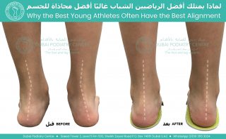
In people with flat feet, the arches collapse more than they should during walking, causing the feet to roll inwards a movement known as overpronation.
As the feet roll inward, the rest of the leg follows.
This can lead to:
- Increased inward rotation of the lower leg
- Changes in knee alignment
- Altered hip mechanics
- Increased stress on muscles, tendons and joints
Rather than one isolated problem, it becomes a chain reaction affecting the entire lower limb.
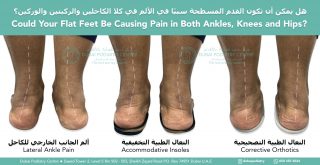
Why Does the Outside of the Ankle Hurt?
Many people expect flat feet to cause pain on the inside of the ankle, but pain on the outside is actually very common.
As the foot collapses inward, the heel tilts and the structures on the outside of the ankle are forced to work harder to stabilise the foot.
This may place excessive strain on:
- The peroneal tendons
- The lateral ankle ligaments
- The muscles that support balance during walking
Over time, these structures become overloaded, resulting in persistent pain on the outside of the ankles.
How Flat Feet Can Lead to Knee Pain
The feet and knees work together with every step.
When the feet overpronate excessively, the shin bone rotates inward. This alters the normal movement of the knee and increases stress across the joint.
Patients commonly experience:
- Pain during walking
- Discomfort when climbing stairs
- Knee fatigue after standing for long periods
- Gradually worsening symptoms with activity
If the underlying foot mechanics are not addressed, the knee continues to compensate with every step.
Why Hip Pain Often Develops
The inward rotation that begins at the foot doesn’t stop at the knee.
It continues into the hips, affecting the muscles that stabilise the pelvis and changing the way the hip joint functions.
This can lead to:
- Hip muscle fatigue
- Pain around the outside of the hips
- Reduced walking endurance
- Difficulty standing for prolonged periods
Many patients are surprised to discover that their hip pain may actually begin with poor foot alignment.
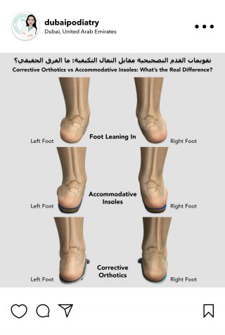
Not All Insoles Work the Same Way
Many patients tell us they have already tried insoles but still have pain.
This is because there are two very different types of insoles.
Accommodative insoles are designed to cushion the feet and improve comfort by redistributing pressure. They can be very helpful for reducing discomfort but do not significantly change foot alignment.
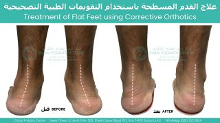
Corrective orthotics, on the other hand, are designed to control excessive foot movement, improve heel position and restore better lower limb alignment. By addressing the underlying biomechanics, they may reduce abnormal loading through the ankles, knees and hips.
The most appropriate option depends on a thorough biomechanical assessment and the specific cause of the patient’s symptoms.
Why a Biomechanical Assessment Is Important
Pain in multiple joints is often treated one area at a time. However, unless the underlying cause is identified, symptoms may continue to return.
A comprehensive biomechanical assessment allows the podiatrist to evaluate:
- Foot posture
- Walking pattern (gait)
- Heel alignment
- Lower limb biomechanics
- Muscle function and joint movement
This helps determine whether abnormal foot mechanics are contributing to pain higher up the body.
Early Treatment Can Prevent Further Problems
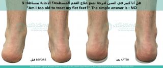
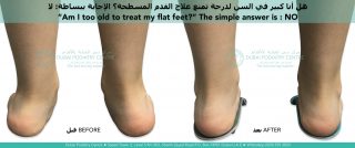
Flat feet do not always require treatment. However, when they are causing pain in the ankles, knees or hips, early intervention can improve function and help prevent further joint overload.
Treatment may include:
- A comprehensive biomechanical assessment
- Custom corrective orthotics where clinically indicated
- Footwear advice
- Stretching and strengthening exercises
- Ongoing monitoring to optimise lower limb alignment
If you have flat feet and are experiencing pain on the outside of both ankles, followed by aching knees and hips, the problem may not be isolated to each joint. Instead, your symptoms could be the result of altered foot mechanics affecting the entire lower limb.
Rather than simply treating the painful area, identifying and correcting the underlying cause can help improve movement, reduce abnormal stress and provide longer-term relief.
At Dubai Podiatry Centre, we assess the body as a connected system because healthy movement starts with a stable foundation.
For more information or to book an appointment please call our clinic +971 4 3435390 or WhatsApp +971 50 3553024