fbpx

Please wait...


BLOG

No more posts

Written by Michelle Champlin BSc Pod., M.Ch.S., S.R., Ch., (UK)

For fungal toenails there are a few treatment options – but which is the best for you? We asked Chief Podiatrist Michelle Champlin of Dubai Podiatry Centre to explain in simple terms the latest treatments – you can also find out more about the signs of fungal toenails.

• There is laser for flat fungal toenails that do not need any corrective treatment for the shape of the nail. This takes a few sessions at least.

• Another effective, fast and immediate treatment is total nail removal, useful if the nail is badly misshapen (including curved or painful ingrowing nails), or if the fungal infection is down into the nail root.

• An alternative option to treat some types of toenail infection is with an oral anti fungal (such as Lamisil tablets), although usually this needs to be taken for 6-9 months and will require an initial and ongoing liver function tests.

• Another way to treat minor superficial fungal nail infection is to use a topical antifungal nail lacquer, such as Curanail. This works nicely on superficial nail fungal infections that can be brought on by covering your toenails excessively with toenail polish or from wearing shoes and socks for a long time during the day, allowing moisture to build up on the top nail plate. This option requires longer term, usually weekly home application with follow up from your Podiatrist to ensure the treatment has resolved the infection fully.

Minimise nail polish use to avoid fungal toenail infections

Visit your Podiatrist to see if laser, total nail removal or medications (superficial or oral) would be the best option for your case. You should always see your Podiatrist if you notice any changes to your toenails, or if you suspect a fungal infection has taken hold. They will be able to check for fungal skin infections also, as this is common alongside fungal toenails. It is important to maintain moisturised, clean toenails as part of an overall toenail care and footcare routine, as dry, damaged or over-pedicured toenails are more at risk of fungal infections.

Contact the foot experts at Dubai Podiatry Centre today in order to get back to healthy toenails that you don’t need to hide.
Call: + 971  4 3435390
WhatsApp: + 971  50 355 3024
info@dubaipodiatry.com
To book online: Click here

بقلم ميشيل شامبلين

هناك عدد قليل من خيارات العلاج لأظافر القدم الفطرية، ولكن ما هو الأفضل بالنسبة لك؟ لقد طلبنا من ميشيل شامبلين، رئيسة أطباء الأقدام في مركز دبي لعلاج الأرجل أن تشرح بعبارات بسيطة أحدث العلاجات – ويمكنك أيضًا معرفة المزيد عن علامات فطريات أظافر القدم.

يوجد ليزر لأظافر القدم الفطرية المسطحة التي لا تحتاج إلى أي علاج تصحيحي لشكل الظفر. وهذا يستغرق بضع جلسات على الأقل.

هناك علاج آخر فعال وسريع وفوري وهو الإزالة الكاملة للأظافر، وهو مفيد إذا كان شكل الظفر مشوهًا بشدة (بما في ذلك الأظافر المنحنية أو المؤلمة)، أو إذا كانت العدوى الفطرية متغلغلة في جذر الظفر.

هناك خيار بديل لعلاج بعض أنواع عدوى أظافر القدم وهو استخدام مضاد للفطريات عن طريق الفم (مثل أقراص لاميسيل)، على الرغم من أنه عادة ما يجب تناوله لمدة 6-9 أشهر وسيتطلب اختبارات وظائف الكبد الأولية والمستمرة.

هناك طريقة أخرى لعلاج عدوى الأظافر الفطرية السطحية البسيطة وهي استخدام طلاء الأظافر الموضعي المضاد للفطريات، مثل Curanail. يعمل هذا بشكل جيد على الالتهابات الفطرية السطحية للأظافر التي يمكن أن تحدث عن طريق تغطية أظافر قدميك بشكل مفرط باستخدام طلاء أظافر القدم أو من ارتداء الأحذية والجوارب لفترة طويلة خلال اليوم، مما يسمح للرطوبة بالتراكم على صفيحة الظفر العلوية. يتطلب هذا الخيار تطبيقًا منزليًا على المدى الطويل وعادة ما يكون أسبوعيًا مع متابعة من طبيب الأقدام للتأكد من أن العلاج قد عالج العدوى بشكل كامل.

التقليل من استخدام طلاء الأظافر لتجنب الإصابة بالفطريات في أظافر القدم

قم بزيارة طبيب الأقدام لمعرفة ما إذا كان الليزر أو إزالة الأظافر بالكامل أو الأدوية (السطحية أو الفموية) هي الخيار الأفضل لحالتك. يجب عليك دائمًا مراجعة طبيب الأقدام الخاص بك إذا لاحظت أي تغييرات في أظافر قدميك، أو إذا كنت تشك في انتشار عدوى فطرية. سيكونون قادرين على التحقق من وجود عدوى جلدية فطرية أيضًا، لأن هذا أمر شائع بجانب أظافر القدم الفطرية. من المهم الحفاظ على أظافر رطبة ونظيفة كجزء من روتين العناية الشامل بأظافر القدمين، حيث أن أظافر القدم الجافة أو التالفة أو المفرطة في العناية بالأظافر تكون أكثر عرضة لخطر الإصابة بالعدوى الفطرية.

تواصل مع خبراء القدم في مركز دبي لعلاج الأرجل اليوم لتتمكن من العودة إلى أظافر صحية لا تحتاج إلى إخفائها.

اتصل ٠٤٣٤٣٥٣٩٠

واتساب: ٠٥٠٣٥٥٣٠٢٤

info@dubaipodiatry.com

للحجز عبر الإنترنت: اضغط هنا


Written by Michelle Champlin BSc Pod., M.Ch.S., S.R., Ch., (UK)
When Is The Best Time To Buy School Shoes?
Whilst you want to avoid the stress and limited shoe choice of a last minute shoe purchase, you don’t want to buy shoes so far in advance that your child has outgrown them before school even starts! Usually, it’s safe to start shoe shopping about now – up to four weeks before school goes back. This way you shouldn’t get caught out by a last minute growth spurt, and also have a wide variety and plenty of time to browse.

Opt for velcro or buckle straps for school shoes
When Do I Need to Buy Another Pair?
The average child’s foot grows about 8mm (a shoe size) per year. A pair of shoes should last for 4-5 months, so get your child’s feet measured again in the New Year. Get measured and shop again sooner if you notice a growth spurt sooner, or the shoes show wear and tear.
How Do I Know if The Shoe is The Right Size?
Shop at a store that has approved, trained shoe fitters. Clarks is one such store – it should be free and no pressure to purchase. Bear in mind that each brand uses different shoe ‘lasts’ or foot models to make their shoes and your child may be different sizes in different brands. Find shoes that have different width fittings – only a third of children have ‘standard’ shoe width feet. Remember to try the shoes on with the socks that your child will wear to school too, and not barefoot to ensure a good fit.
What Type Of Shoe Should You Buy?
Check out our guide to school shoes here. For younger kids who may struggle with tying laces, opt for Velcro fasteners, or buckles. Older kids can choose lace up styles, or mary jane style straps across the foot. It’s important that shoes are fastened securely to the foot to encourage active play and even for older children; a shoe held securely to the foot is preferable to slip on styles such as ballet pumps. Slip on styles can encourage biomechanical issues such as over-pronation (fallen arches) and toe clawing. They also don’t provide enough shock absorption in the sole. A slight heel of a couple of centimeters is also a good feature, a rubberized non-slip sole that is shock absorbing yet bendable at the toe. A stiff heel counter, plenty of room at the toe box so as not to squash the growing toes and forefoot. Natural material such as leather or breathable man made fabrics such as Gore-Tex to stop excessive sweat build up and possible fungal infections. A removable shoe liner too if your child wears orthotics (custom insoles) prescribed by your Podiatrist to correct issues such as over-pronation / flat feet.
Adults aim to take 10,000 steps a day to keep fit but kids can take 18,000 on the average school day. School shoes are much more than a fashion statement, as children’s feet do not stop forming and growing until they are around 18 years old. Wearing ill-fitting school shoes in childhood can cause permanent structural damage to the feet with life long consequences, and exacerbate biomechanical issues such as over-pronation. Children spend over 35 hours a week in these shoes, so they must be durable, high quality and well fitting.
Why Would My Child Need To See A Podiatrist?
It is vital that all children have their feet screened by a Podiatrist as routine, normally when they start school for the first time (age 5-6) and again as advised by the Podiatrist, just as they go for their first dental check up. Dubai Podiatry Centre, led by Chief Podiatrist Michelle Champlin, carries out a popular Foot Health Screening Program at the Schools in Dubai, but don’t worry if yours missed it – just contact us and pop into our clinic for a friendly and fun appointment before school starts back. Getting an early foot check reduces the chances of foot problems in later life, gives and opportunity to address any issues early and establishes an understanding of good foot health habits early.
You may not know if there is anything going on with your kid’s feet, until they start complaining of pain or are tripping a lot. The Podiatrist can check your child’s alignment, advise on footwear, as well as catch any early fungal or verruca skin infections commonly passed around.
For your recommended kid’s foot check up before the school term starts, contact Dubai Podiatry Centre on +971 4 3435390


العودة إلى المدرسة – الإجابة على أسئلتك المتعلقة بالأحذية المدرسية

بقلم ميشيل شامبلين

ما هو أفضل وقت لشراء الأحذية المدرسية؟

على الرغم من أنك ترغب في تجنب التوتر ومحدودية اختيار الأحذية عند شراء حذاء في اللحظة الأخيرة، إلا أنك لا ترغب في شراء الأحذية مقدمًا حتى أن طفلك قد تجاوزها حتى قبل أن يبدأ المدرسة! عادةً ما يكون من الآمن البدء في شراء الأحذية الآن – ما يصل إلى أربعة أسابيع قبل عودة المدرسة. بهذه الطريقة، لن تنزعج من طفرة النمو في اللحظة الأخيرة، وسيكون لديك أيضًا مجموعة واسعة ومتسع من الوقت للتصفح.

اختر أشرطة الفيلكرو أو الإبزيم للأحذية المدرسية

متى أحتاج إلى شراء زوج آخر؟

ينمو متوسط قدم الطفل حوالي ٨ ملم (حجم الحذاء) سنويًا. يجب أن يستمر زوج الأحذية لمدة ٤-٥ أشهر، لذا قم بقياس قدم طفلك مرة أخرى في العام الجديد. قم بالقياس والتسوق مرة أخرى عاجلاً إذا لاحظت طفرة في النمو عاجلاً، أو إذا ظهرت الأحذية على شكل تآكل.

كيف أعرف إذا كان الحذاء هو الحجم المناسب؟

تسوق في متجر معتمد ومدرب لتركيب الأحذية. كلاركس هو أحد هذه المتاجر – يجب أن يكون مجانيًا ولا يوجد أي ضغط للشراء. ضع في اعتبارك أن كل علامة تجارية تستخدم نماذج مختلفة من الأحذية أو نماذج القدم لصنع أحذيتها وقد يكون لطفلك أحجام مختلفة في ماركات مختلفة. ابحث عن أحذية ذات مقاسات عرض مختلفة – ثلث الأطفال فقط لديهم أقدام بعرض حذاء “قياسي”. تذكر أن تجرب الحذاء مع الجوارب التي سيرتديها طفلك في المدرسة أيضًا، وليس حافي القدمين لضمان ملاءمة جيدة.

ما نوع الحذاء الذي يجب أن تشتريه؟

تحقق من دليلنا للأحذية المدرسية هنا. بالنسبة للأطفال الأصغر سنًا الذين قد يواجهون صعوبة في ربط الأربطة، اختر مثبتات الفيلكرو أو الأبازيم. يمكن للأطفال الأكبر سنًا اختيار أنماط بأربطة أو أشرطة بنمط ماري جين على القدم. من المهم أن يتم تثبيت الأحذية بشكل آمن على القدم لتشجيع اللعب النشط وحتى للأطفال الأكبر سنًا؛ من الأفضل أن يتم تثبيت الحذاء بشكل آمن على القدم بدلاً من الانزلاق على أنماط مثل أحذية الباليه. يمكن أن تؤدي الأنماط سهلة الارتداء إلى تشجيع المشكلات الميكانيكية الحيوية مثل الإفراط في الكب (الأقواس الساقطة) ومخالب أصابع القدم. كما أنها لا توفر ما يكفي من امتصاص الصدمات في النعل. يعد الكعب الخفيف الذي يبلغ بضعة سنتيمترات أيضًا ميزة جيدة، وهو نعل مطاطي غير قابل للانزلاق يمتص الصدمات ولكنه قابل للانحناء عند إصبع القدم. كعب صلب، مساحة كبيرة في صندوق أصابع القدم حتى لا تسحق أصابع القدم ومقدمة القدم النامية. المواد الطبيعية مثل الجلد أو الأقمشة المصنوعة يدويًا مثل Gore-Tex لوقف تراكم العرق الزائد والالتهابات الفطرية المحتملة. بطانة حذاء قابلة للإزالة أيضًا إذا كان طفلك يرتدي أجهزة تقويم العظام (نعال داخلية مخصصة) يصفها طبيب الأقدام الخاص بك لتصحيح مشكلات مثل الإفراط في الكب / القدم المسطحة.

يهدف البالغون إلى اتخاذ ١٠٠٠٠ خطوة يوميًا للحفاظ على لياقتهم البدنية، ولكن يمكن للأطفال القيام بـ ١٨٠٠٠ خطوة في اليوم الدراسي العادي. الأحذية المدرسية هي أكثر من مجرد موضة، حيث أن أقدام الأطفال لا تتوقف عن التشكل والنمو حتى يبلغوا حوالي ١٨ عامًا. يمكن أن يؤدي ارتداء أحذية مدرسية غير مناسبة في مرحلة الطفولة إلى حدوث ضرر هيكلي دائم للقدمين مع عواقب مدى الحياة، كما يؤدي إلى تفاقم المشكلات الميكانيكية الحيوية مثل الإفراط في الكب. يقضي الأطفال أكثر من ٣٥ ساعة أسبوعيًا في ارتداء هذه الأحذية، لذا يجب أن تكون متينة وعالية الجودة وملائمة تمامًا.

لماذا يحتاج طفلي لرؤية طبيب الأقدام؟

من الضروري أن يتم فحص أقدام جميع الأطفال من قبل طبيب الأقدام بشكل روتيني، عادة عندما يبدأون المدرسة لأول مرة (من سن ٥ إلى ٦ سنوات) ومرة أخرى حسب نصيحة طبيب الأقدام، تمامًا كما يذهبون لإجراء فحص أسنانهم الأول. ينفذ مركز دبي لطب الأرجل، بقيادة كبير أطباء الأقدام ميشيل شامبلين، برنامجًا شائعًا لفحص صحة القدم في المدارس في دبي، ولكن لا تقلق إذا فاتتك هذه البرامج – ما عليك سوى الاتصال بنا والدخول إلى عيادتنا للحصول على موعد ودود وممتع قبل ذلك. تبدأ المدرسة مرة أخرى. إن إجراء فحص مبكر للقدم يقلل من فرص حدوث مشاكل في القدم في وقت لاحق من الحياة، ويمنح الفرصة لمعالجة أي مشاكل في وقت مبكر ويؤسس لفهم العادات الصحية الجيدة للقدم في وقت مبكر.

قد لا تعرفين ما إذا كان هناك أي شيء يحدث بقدمي طفلك، حتى يبدأ في الشكوى من الألم أو يتعثر كثيرًا. يمكن لطبيب الأقدام التحقق من استقامة طفلك، وتقديم المشورة بشأن الأحذية، بالإضافة إلى اكتشاف أي التهابات جلدية فطرية أو ثؤلولية مبكرة تنتشر عادة حوله.

لإجراء فحص قدم طفلك الموصى به قبل بدء الفصل الدراسي، اتصل بمركز دبي للعناية بالاقدام على الرقم ٠٤٣٤٣٥٣٩٠  


Written by Michelle Champlin BSc Pod., M.Ch.S., S.R., Ch., (UK)
At Dubai Podiatry Centre, our Podiatrists frequently see children with many different foot issues. This can range from skin infections such as verrucas and fungal infections to issues affecting their bones, tendons, muscles or ligaments such as hypermobility, flat feet, or Sever’s Disease. Ingrown toenails are particularly common amongst teenagers. Symptoms of foot ailments can be subtle, and young children may not know how to express their foot problem.
Sometimes it can be difficult to tell that your child has a foot or toenail problem. They might be shy, not aware that they have a problem or that it can be fixed. Foot issues in children must be addressed as priority, as their foot’s bones and structure are still developing, with untreated ailments potentially having life long impact.

A child becoming reluctant to take part in sports can be a sign of foot problems
Chief Podiatrist Michelle Champlin outlines five common signs that your child may have a foot or leg problem:
• You notice that your child seems ‘clumsy’ – tripping or falling a lot – this may be caused by in-toeing, neuromuscular or balance challenges.
• Your child tires easily and is reluctant to walk more than short distances. With biomechanical issues such as over-pronation or flat feet, the feet and legs have to work harder and are less efficient, thus muscles tire quickly. If your child is lagging behind in the playground or races, foot and leg fatigue may be the cause, related to a biomechanical issue.
• Your child complains of pain. No form of foot or leg pain is normal in a child. Pain that lasts beyond a few days should be investigated.
• Your child becomes reluctant to take part in PE, games or physical activity that they used to enjoy – particularly weight bearing games such as football, running or tennis. Conditions such as Sever’s Disease are prone to very active, sporty children aged between 8 and 14.
• Your child doesn’t want to show you their feet. We encourage children to get used to their parents inspecting their feet from a young age and developing good foot hygiene, care and footwear. Teenagers particularly can suffer from painful ingrowing toenails during their growth spurts and can sometimes try to self manage at home. Ingrowing toenails can be easily and permanently resolved – at Dubai Podiatry Centre we treat patients’ feet as gently as our own children’s and understand that coming to see the Podiatrist can be a nervous time for children.
If your child is exhibiting any of the above symptoms, or if you would like a routine foot health check as we advise all children from the age of 5/6 years old, make an appointment today on +971 4 3435390.


بقلم ميشيل شامبلين

في مركز دبي لعلاج الأقدام، كثيرًا ما يقوم أطباء الأقدام لدينا بفحص الأطفال الذين يعانون من العديد من مشاكل القدم المختلفة. يمكن أن يتراوح هذا من الالتهابات الجلدية مثل الثآليل والالتهابات الفطرية إلى المشكلات التي تؤثر على العظام أو الأوتار أو العضلات أو الأربطة مثل فرط الحركة أو الأقدام المسطحة أو مرض سيفر. أظافر القدم الناشبة شائعة بشكل خاص بين المراهقين. يمكن أن تكون أعراض أمراض القدم خفية، وقد لا يعرف الأطفال الصغار كيفية التعبير عن مشكلة قدمهم.

في بعض الأحيان قد يكون من الصعب معرفة أن طفلك يعاني من مشكلة في القدم أو أظافر القدم. قد يكونون خجولين، ولا يدركون أن لديهم مشكلة أو أنه يمكن حلها. يجب معالجة مشاكل القدم لدى الأطفال كأولوية، حيث أن عظام قدمهم وبنيتها لا تزال في طور النمو، ومن المحتمل أن يكون للأمراض غير المعالجة تأثيرًا مدى الحياة.

قد يكون تردد الطفل في ممارسة الرياضة علامة على وجود مشاكل في القدم

توضح رئيسة أطباء الأقدام ميشيل شامبلين خمس علامات شائعة تشير إلى أن طفلك قد يعاني من مشكلة في القدم أو الساق:

لاحظت أن طفلك يبدو “أخرقًا” – يتعثر أو يسقط كثيرًا – قد يكون سبب ذلك تحديات عصبية عضلية أو تحديات في التوازن.

يتعب طفلك بسهولة ويتردد في المشي لأكثر من مسافات قصيرة. مع المشكلات الميكانيكية الحيوية مثل الإفراط في الكب أو القدم المسطحة، يجب على القدمين والساقين العمل بجهد أكبر وتكون أقل كفاءة، وبالتالي تتعب العضلات بسرعة. إذا كان طفلك متخلفًا في الملعب أو السباقات، فقد يكون تعب القدم والساق هو السبب، وهو ما يتعلق بمشكلة ميكانيكية حيوية.

طفلك يشكو من الألم. لا يوجد أي شكل من أشكال الألم في القدم أو الساق أمر طبيعي عند الطفل. يجب التحقق من الألم الذي يستمر لأكثر من بضعة أيام.

يصبح طفلك متردداً في المشاركة في التربية البدنية أو الألعاب أو الأنشطة البدنية التي كان يستمتع بها – وخاصة ألعاب حمل الأثقال مثل كرة القدم أو الجري أو التنس. تكون الحالات مثل مرض سيفر عرضة للأطفال النشطين للغاية والرياضيين الذين تتراوح أعمارهم بين ٨ و١٤ عامًا.

لا يريد طفلك أن يظهر لك قدميه. نحن نشجع الأطفال على الاعتياد على قيام والديهم بفحص أقدامهم منذ الصغر وتطوير النظافة الجيدة للقدمين والعناية بها والأحذية. يمكن أن يعاني المراهقون على وجه الخصوص من ألم في أظافر القدمين أثناء طفرات نموهم ويمكنهم أحيانًا محاولة التحكم بأنفسهم في المنزل. يمكن حل مشكلة أظافر القدم الغارزة بسهولة وبشكل دائم – في مركز دبي لعلاج الأقدام، نتعامل مع أقدام المرضى بلطف مثل أقدام أطفالنا وندرك أن القدوم لرؤية طبيب الأقدام يمكن أن يكون وقتًا عصبيًا للأطفال.

إذا كان طفلك يعاني من أي من الأعراض المذكورة أعلاه، أو إذا كنت ترغب في إجراء فحص روتيني لصحة القدم كما ننصح جميع الأطفال من سن ٥/٦ سنوات

حدد موعدًا اليوم على الرقم ٠٤٣٤٣٥٣٩٠


Your feet support the entire weight of your body and having healthy, pain free feet allows you to go anywhere and do anything without worrying about debilitating foot pain.

Your feet are made up of a complex network of bones, muscles, tendons, ligaments, nerves, arteries and veins. To narrow down the cause of foot pain, let’s look at the areas of the foot you might experience pain. One of the most common foot areas for pain is the heel.

Plantar fasciitis

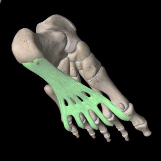
This is the most common cause of heel pain, reports Chief Podiatrist Michelle Champlin. The plantar fascia is an extremely tough band of fibrous tissue that connects your heel bone (calcaneum) to the front of your foot, just behind your toes.

Plantar fasciitis is the most common cause of heel pain

The plantar fascia is similar to an emergency brake for your foot and can become irritated or inflamed when overused. This can happen when the muscles of your foot, particularly those forming the long arch of your foot, are untoned and not engaging properly when you run or walk. You may notice that your foot looks ‘flat’ due to the collapsed long arch.

Plantar fasciitis due to an underlying biomechanical cause can be assessed and diagnosed by your Podiatrist. Treatment may involve wearing custom corrective orthotics inside your work or leisure shoes, stretching and strengthening exercises and pain relieving NSAIDs. Dubai Podiatry Centre is the region’s only clinic which provides a one-stop service by qualified Podiatrists from assessing the patient, through to taking a mold of the patient’s feet, prescribing the correct orthotic, making them in-clinic in their own lab and then fitting them, as well as closely monitoring progress and making any ongoing adjustments required as your foot shape changes and foot pain reduces.

Repeated use of steroid injections should be avoided due to their reduction of the fatty padding in the heel, which will worsen the problem in the long run.

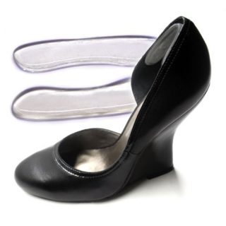
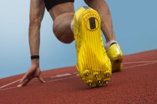
Fallen arches are not the only underlying cause of plantar fasciitis. Other common causes include very high arched foot types (pes cavus), sudden increase in exercise regime, wearing thin soled shoes such as ballet pumps or flip flops, a difference in leg lengths, short Achilles tendon, or an increase in bodyweight.

All of our bodies are unique, with different stresses and strains, so it’s important to see a qualified Podiatrist for heel pain to ensure treatment for the root cause and long-term resolution of the pain.

Other causes of heel pain can include heel spurs, stone bruises or Achilles tendonitis. Generalized foot pain, especially burning or tingling, should always be investigated thoroughly by your GP or Podiatrist, especially if your are diabetic. These can be signs of neuropathy, or nerve damage, and can signal that changes to your medication, diet or exercise routine are required.

For appointment, contact us:
Call: + 971  4 3435390
WhatsApp: + 971  50 355 3024
info@dubaipodiatry.com
To book online: Click here

ألم الكعب: هل هو التهاب اللفافة الأخمصية؟

تدعم قدميك وزن جسمك بالكامل، كما أن الحصول على أقدام صحية وخالية من الألم يسمح لك بالذهاب إلى أي مكان وفعل أي شيء دون القلق بشأن آلام القدم المنهكة.

تتكون قدميك من شبكة معقدة من العظام والعضلات والأوتار والأربطة والأعصاب والشرايين والأوردة. لتضييق نطاق سبب آلام القدم، دعونا نلقي نظرة على مناطق القدم التي قد تعاني من الألم. أحد أكثر مناطق القدم شيوعًا للألم هو الكعب

التهاب اللفافة الأخمصية

هذا هو السبب الأكثر شيوعًا لألم الكعب، وفقًا لتقارير رئيسة أطباء الأقدام ميشيل شامبلين. اللفافة الأخمصية عبارة عن شريط قوي للغاية من الأنسجة الليفية التي تربط عظم الكعب (العقبي) بمقدمة قدمك، خلف أصابع قدميك مباشرةً

التهاب اللفافة الأخمصية هو السبب الأكثر شيوعًا لألم الكعب

تشبه اللفافة الأخمصية فرامل الطوارئ لقدمك ويمكن أن تصبح متهيجة أو ملتهبة عند الإفراط في استخدامها. يمكن أن يحدث هذا عندما تكون عضلات قدمك، خاصة تلك التي تشكل القوس الطويل لقدمك، غير متماسكة ولا تعمل بشكل صحيح عند الجري أو المشي. قد تلاحظ أن قدمك تبدو “مسطحة” بسبب القوس الطويل المنهار

يمكن تقييم وتشخيص التهاب اللفافة الأخمصية الناتج عن سبب ميكانيكي حيوي أساسي من قبل طبيب الأقدام الخاص بك. قد يشمل العلاج ارتداء أجهزة تقويم العظام التصحيحية المخصصة داخل أحذية العمل أو الترفيه، وتمارين التمدد والتقوية ومضادات الالتهاب غير الستيروئيدية المخففة للألم. مركز دبي لعلاج الأرجل هو العيادة الوحيدة في المنطقة التي تقدم خدمة شاملة من قبل أطباء الأقدام المؤهلين بدءًا من تقييم المريض وحتى أخذ قالب لقدم المريض ووصف جهاز تقويم العظام الصحيح وجعله داخل العيادة في مختبره الخاص ومن ثم تركيبه. لهم، بالإضافة إلى مراقبة التقدم عن كثب وإجراء أي تعديلات مستمرة مطلوبة مع تغير شكل قدمك وتقليل آلام القدم

يجب تجنب الاستخدام المتكرر لحقن الستيرويد لأنها تقلل من الحشوة الدهنية في الكعب، مما يؤدي إلى تفاقم المشكلة على المدى الطويل

الأقواس المتساقطة ليست السبب الوحيد الكامن وراء التهاب اللفافة الأخمصية. تشمل الأسباب الشائعة الأخرى أنواع القدم المقوسة جدًا (pes cavus)، أو الزيادة المفاجئة في نظام التمارين الرياضية، أو ارتداء أحذية ذات نعل رفيع مثل أحذية الباليه أو النعال، أو الاختلاف في أطوال الساق، أو قصر وتر العرقوب، أو زيادة في وزن الجسم

جميع أجسامنا فريدة من نوعها، مع ضغوطات وتوترات مختلفة، لذلك من المهم رؤية طبيب أقدام مؤهل لعلاج آلام الكعب لضمان علاج السبب الجذري للألم وحله على المدى الطويل

يمكن أن تشمل الأسباب الأخرى لألم الكعب نتوءات الكعب أو الكدمات الحجرية أو التهاب وتر العرقوب. يجب دائمًا فحص آلام القدم العامة، وخاصة الحرق أو الوخز، بدقة من قبل طبيبك أو طبيب الأقدام، خاصة إذا كنت مصابًا بالسكري. يمكن أن تكون هذه علامات على الاعتلال العصبي أو تلف الأعصاب، ويمكن أن تشير إلى ضرورة إجراء تغييرات على الدواء أو النظام الغذائي أو روتين التمارين الرياضية

‎لحجز موعد، تواصل معنا: الهاتف: ٠٤٣٤٣٥٣٩٠
واتساب: ٠٥٠٣٥٥٣٠٢٤
البريد الإلكتروني: info@dubaipodiatry.com
لحجز موعد عبر الإنترنت: اضغط هنا


Written by Michelle Champlin BSc Pod., M.Ch.S., S.R., Ch., (UK)
Sudden onset of heel pain can be due to standing on a sharp object whilst barefoot, such as a stone or piece of Lego. Ouch! It can also be caused by a sudden impact such as landing awkwardly if you jump off a height. The pain can be excruciating at first and even form a deep bruise on the soft tissue and fatty padding of your heel – called a ‘stone bruise’.
The pain can feel like a constant discomfort, as if there is a small stone or pebble in your shoe. Persistent heel pain should always be checked out by your GP or Podiatrist, to ensure that you have not fractured your heel bone (calcaneum). You can read more about heel fractures here.
Your Podiatrist will be able to diagnose what’s causing your heel pain, by examining your foot, taking a thorough history, assessing how you stand and walk and perhaps ordering an x-ray or MRI. Here’s some of the differences between a heel bruise and plantar fasciitis, which is the leading cause of heel pain:
• A bruise will feel like a constant pain when walking – plantar fasciitis pain tends to be at its worst on the first few steps in the morning or after resting.
• You may see evidence of blue/black bruising on your heel with a bruise – no outward signs may be present with plantar fasciitis.
• Plantar fasciitis tends to be an overuse injury, linked with an underlying biomechanical issue such as flat feet or high arches.
• A heel bruise tends to be due to a sudden trauma such as standing on an object or jumping onto the heel.

Try resting, elevating and icing a heel bruise
Stone bruises usually resolve with icing to the area, resting and elevating the foot as much as possible and wearing soft, cushioned shoes with plenty of shock absorption. Severe bruising resistant to healing may require the use of an aircast to keep weight off of the heel.
See your Podiatrist for advice on pain relief medication such as ibuprofen, footwear assessment and advice or possibly in-shoe padding or orthotics (medical insoles) to offload pressure or correct your gait if required. They will also be able to diagnose whether the pain is due to plantar fasciitis, bruise, heel spur, bone fracture or Achilles tendonitis as all can cause heel pain. Sometimes, you may also feel a stone bruise to your forefoot, or ball of foot. Again, these symptoms of metatarsalgia might be due to a simple bruise, or signify a metatarsal stress fracture or transverse arch collapse. Your Podiatrist will be able to correctly spot it and provide the right treatment.
It is also important to ensure that you don’t have a foreign object embedded in your heel or foot either, such as small piece of glass or broken needle – your Podiatrist or Doctor can easily rule this out with examination and/or x-ray.
Contact the UAE’s foot and leg experts on +971 4 3435390 if you are experiencing any form of heel pain, whether sudden or gradual.


بقلم ميشيل شامبلين

يمكن أن يكون ظهور ألم الكعب المفاجئ نتيجة للوقوف على جسم حاد وأنت حافي القدمين، مثل حجر أو قطعة ليغو. أوه! ويمكن أيضًا أن يكون سببه تأثير مفاجئ مثل الهبوط بشكل غريب إذا قفزت من ارتفاع. يمكن أن يكون الألم مؤلمًا في البداية وقد يشكل كدمة عميقة على الأنسجة الرخوة والبطانة الدهنية لكعبك – تسمى “الكدمة الحجرية“.

قد يبدو الألم كأنه انزعاج مستمر، كما لو كان هناك حجر صغير أو حصاة في حذائك. يجب دائمًا فحص ألم الكعب المستمر من قبل طبيبك العام أو طبيب الأقدام للتأكد من عدم تعرضك لكسر في عظم الكعب (العظم). يمكنك قراءة المزيد عن كسور الكعب هنا.

سيكون طبيب الأقدام الخاص بك قادرًا على تشخيص سبب ألم كعبك، عن طريق فحص قدمك، وأخذ تاريخك الدقيق، وتقييم كيفية وقوفك ومشيك، وربما طلب تصوير بالأشعة السينية أو التصوير بالرنين المغناطيسي. فيما يلي بعض الاختلافات بين كدمة الكعب والتهاب اللفافة الأخمصية، وهو السبب الرئيسي لألم الكعب:

ستشعر بالكدمة وكأنها ألم مستمر عند المشي – يميل ألم التهاب اللفافة الأخمصية إلى أن يكون في أسوأ حالاته في الخطوات القليلة الأولى في الصباح أو بعد الراحة.

قد ترى دليلاً على وجود كدمة زرقاء/سوداء على كعبك مع كدمة – قد لا توجد علامات خارجية مع التهاب اللفافة الأخمصية.

يميل التهاب اللفافة الأخمصية إلى الإصابة الناتجة عن الإفراط في الاستخدام، ويرتبط بمشكلة ميكانيكية حيوية كامنة مثل القدم المسطحة أو الأقواس المرتفعة.

تميل كدمة الكعب إلى أن تكون نتيجة لصدمة مفاجئة مثل الوقوف على جسم ما أو القفز على الكعب.

حاول الراحة والرفع ووضع الثلج على كدمة الكعب

عادة ما يتم حل الكدمات الحجرية بوضع الثلج على المنطقة، مع الراحة ورفع القدم قدر الإمكان وارتداء أحذية ناعمة ومبطنة مع الكثير من امتصاص الصدمات. قد تتطلب الكدمات الشديدة المقاومة للشفاء استخدام جهاز بث هوائي للحفاظ على الوزن بعيدًا عن الكعب.

قم بزيارة طبيب الأقدام الخاص بك للحصول على المشورة بشأن أدوية تخفيف الألم مثل الأيبوبروفين، وتقييم الأحذية والمشورة أو ربما حشوة الحذاء أو تقويم العظام (النعال الطبية) لتخفيف الضغط أو تصحيح مشيتك إذا لزم الأمر. سيكونون أيضًا قادرين على تشخيص ما إذا كان الألم ناتجًا عن التهاب اللفافة الأخمصية أو الكدمة أو مهماز الكعب أو كسر العظام أو التهاب وتر العرقوب حيث أن جميعها يمكن أن تسبب ألم الكعب. في بعض الأحيان، قد تشعر أيضًا بكدمة حجرية في مقدمة قدمك، أو كرة قدمك. مرة أخرى، قد تكون أعراض ألم مشط القدم هذه ناتجة عن كدمة بسيطة، أو تشير إلى كسر في مشط القدم أو انهيار القوس المستعرض. سيكون طبيب الأقدام الخاص بك قادرًا على اكتشافه بشكل صحيح وتقديم العلاج المناسب.

من المهم أيضًا التأكد من عدم وجود جسم غريب مغروس في كعبك أو قدمك أيضًا، مثل قطعة صغيرة من الزجاج أو إبرة مكسورة – يمكن لطبيب الأقدام أو الطبيب استبعاد ذلك بسهولة من خلال الفحص و/أو الأشعة السينية. .

تواصل مع خبراء القدم والساق في الإمارات العربية المتحدة على الرقم ٠٤٣٤٣٥٣٩٠ إذا كنت تعاني من أي شكل من أشكال آلام الكعب، سواء كانت مفاجئة أو تدريجية.


Written by Michelle Champlin BSc Pod., M.Ch.S., S.R., Ch., (UK)
A heel spur is a bony growth on the bottom of your calcaneum (heel bone). The reasons for heel spur formation are still being researched, though there is evidence to support both spur formation due to excess pulling on the plantar fascia band where it connects to the heel bone, and also due to vertical forces i.e. downward pressure on the heel from a lot of standing or walking. Research also shows that heel spurs are almost unheard of in those under 20 and still rare in those under 40. They tend to form as we enter our 50s. Women also tend to be more prone to heel spurs than men.
Generally, heel spurs are thought to be caused by the other muscles, tendons and ligaments of the foot not working together as they should, such as a collapsed long arch causing the foot to over-pronate and roll in, or plantar fasciitis causing excess strain on the plantar fascia band.

Heel spurs can be treated non-surgically with orthotics
Other risk factors for developing heel spurs include:

• Sports activities involving a lot of jumping or running
• Increase in body weight causing excess force through the feet
• Footwear with insufficient cushioning or support
More on plantar fasciitis and its risk factors can be found in our recent blog here.
Having a heel spur does not necessarily mean you will have symptoms. Many people have heel spurs with no symptoms. However, some spurs do cause intermittent or chronic ongoing pain. It’s generally not the bony spur itself but the irritation and inflammation of the soft tissue around it, causing pain when weight bearing. The pain tends to be worse in the morning or when first standing up, with the sharp pain turning into a dull throb.
If you experience ongoing heel pain, you should make an appointment with your Podiatrist, who will thoroughly investigate and correctly diagnose the source – whether heel bone fracture, plantar fasciitis, stone bruise or more. For heel spurs, you may be advised:
• Shock absorbing footwear
• Stretching exercises for the morning
• Custom orthotics
Avoid repeated steroid injections, as they can worsen the condition in the long term, due to the ‘melting’ of your good fatty padding on the heel.
For any type of heel or foot pain, make an appointment with the UK qualified Podiatrists at Dubai Podiatry Centre on +971 4 3435390.


بقلم ميشيل شامبلين

مهماز الكعب هو نمو عظمي في الجزء السفلي من عظمة الكعب (عظم الكعب). لا تزال أسباب تشكل مهماز الكعب قيد البحث، على الرغم من وجود أدلة تدعم تكوين مهماز الكعب بسبب السحب الزائد على شريط اللفافة الأخمصية حيث يتصل بعظم الكعب، وأيضًا بسبب القوى العمودية، أي الضغط الهبوطي على الكعب من الكثير من الوقوف أو المشي. تظهر الأبحاث أيضًا أن مهماز الكعب لم يُسمع بها تقريبًا لدى الأشخاص الذين تقل أعمارهم عن 20 عامًا، ولا تزال نادرة لدى الأشخاص الذين تقل أعمارهم عن ٤٠ عامًا. وتميل إلى التشكل مع دخولنا الخمسينات من العمر. تميل النساء أيضًا إلى أن تكون أكثر عرضة لمسمار الكعب من الرجال.

بشكل عام، يُعتقد أن مهماز الكعب ناتجة عن عدم عمل العضلات والأوتار والأربطة الأخرى في القدم معًا كما ينبغي، مثل القوس الطويل المنهار الذي يتسبب في الإفراط في كب القدم وتدحرجها، أو التهاب اللفافة الأخمصية الذي يسبب إجهادًا زائدًا على شريط اللفافة الأخمصية.

يمكن علاج نتوءات الكعب بشكل غير جراحي باستخدام تقويم العظام

تشمل عوامل الخطر الأخرى لتطوير مهماز الكعب ما يلي:

الأنشطة الرياضية التي تتضمن الكثير من القفز أو الجري

زيادة وزن الجسم مما يسبب قوة زائدة عبر القدمين

الأحذية التي لا تحتوي على توسيد أو دعم كافٍ

يمكن العثور على المزيد عن التهاب اللفافة الأخمصية وعوامل الخطر المرتبطة به في مدونتنا الأخيرة هنا.

وجود مهماز الكعب لا يعني بالضرورة أنه سيكون لديك أعراض. يعاني العديد من الأشخاص من نتوءات الكعب دون ظهور أي أعراض. ومع ذلك، فإن بعض المهمازات تسبب ألمًا متقطعًا أو مزمنًا مستمرًا. لا يتعلق الأمر عمومًا بالحافز العظمي نفسه، بل هو تهيج والتهاب الأنسجة الرخوة المحيطة به، مما يسبب الألم عند حمل الوزن. يميل الألم إلى أن يكون أسوأ في الصباح أو عند الوقوف لأول مرة، مع تحول الألم الحاد إلى خفقان خفيف.

إذا كنت تعاني من ألم مستمر في الكعب، فيجب عليك تحديد موعد مع طبيب الأقدام الخاص بك، والذي سيقوم بإجراء فحص شامل وتشخيص المصدر بشكل صحيح – سواء كان كسر في عظم الكعب، أو التهاب اللفافة الأخمصية، أو كدمة حجرية أو أكثر. بالنسبة لمهماز الكعب، قد يُنصح بما يلي:

أحذية ممتصة للصدمات

تمارين التمدد في الصباح

أجهزة تقويم العظام المخصصة

تجنب تكرار حقن الستيرويد، لأنها يمكن أن تؤدي إلى تفاقم الحالة على المدى الطويل، بسبب “ذوبان” الحشوة الدهنية الجيدة على الكعب.

لأي نوع من آلام الكعب أو القدم، حدد موعدًا مع أطباء الأقدام المؤهلين من المملكة المتحدة في مركز دبي للعناية بالأقدام على الرقم ٠٤٣٤٣٥٣٩٠.


Written by Michelle Champlin BSc Pod., M.Ch.S., S.R., Ch., (UK)
Your calcaneum, or heel bone is the first part of your body to hit the ground while walking – the ‘heel strike’. Fractures, or breaks to the bone itself are a more unusual cause of heel pain. Other more common causes are plantar fasciitis, Achilles tendonitis and heel spurs, or Sever’s Disease in children.
The heel bone is like a hard boiled egg – it has a thin but hard outer ‘shell’ and a more spongy, softer bone within. The main cause of heel fractures are accidents such as falling onto your heels from a height such as a ladder, or impact during a car crash. Sometimes the heel bone can suffer from stress fractures due to repeated impact over time.

The calcaneum forms the ‘sub-talar’ joint with the talus and cuboid bones
The heel bone connects to two other bones – the talus and the cuboid, as you can see in the picture. These three bones form the sub-talar joint, which controls much of your foot during motion from walking to running and can be a main cause of ‘flat feet’ when the joint allows the foot to ‘over-pronate.’
When serious injuries break not just the heel bone, but also involve this sub-talar joint and it’s ligaments, muscles or tendons, this can be a more complex and difficult injury to mend.
The signs and symptoms of a heel bone fracture depend on where the bone is fractured, how it happened and how severe it is. As with most bone fractures, you will notice pain and swelling to the heel area, with immediate pain if it was a traumatic injury.
To diagnose the injury, the Podiatrist, GP or orthopaedic foot surgeon will take a full history, including events that led up to the pain to understand the mechanism of the injury, physically examine your foot and ankle and order x-rays.

X-ray image of a fractured calcaneum
Treatment for fractured heel bones will depend on the type of fracture and severity. This will indicate whether or not surgery is required.
Non-surgical treatments can include:
• Rest, Ice, Compression and Elevation
• Immobilization and weight being taken off the heel, using a device such as an Aircast
Following healing of the fracture, you may be advised custom orthotic shoe inserts that may be required to correct foot posture and alignment and to tone up underused ligaments and muscles following immobilization. This type of rehabilitation will also likely be advised after surgery too for more serious fractures. Any foot or ankle injury, even ankle sprains from years ago, can have long lasting detrimental effect on your foot and ankle posture (including stiffness, arthritis and pain), so it is important to have any form of heel, foot or ankle pain thoroughly investigated by your Podiatrist.
If you suspect a fractured heel bone, or if you have recovered from a foot or ankle sprain or fracture it is advisable to have your alignment reviewed by a Podiatrist in order to avoid longer-term complications.
Appointments with the leading UK qualified Podiatrists at Dubai Podiatry Centre, led by Chief Podiatrist Michelle Champlin and based on Sheikh Zayed Road, can be made by contacting +971 4 3435390. The Clinic is conveniently located near DIFC Metro station and is open every day except Fridays. If driving, Dubai Podiatry Centre is also listed on the Dubai map app for android and iPhones, Makani.


بقلم ميشيل شامبلين

عظم الكعب أو عظم الكعب هو الجزء الأول من جسمك الذي يضرب الأرض أثناء المشي – “ضربة الكعب”. تعتبر الكسور أو الكسر في العظم نفسه سببًا غير عادي لألم الكعب. الأسباب الأخرى الأكثر شيوعًا هي التهاب اللفافة الأخمصية، أو التهاب وتر العرقوب، أو مرض سيفر عند الأطفال.

يشبه عظم الكعب البيضة المسلوقة – فهو يحتوي على “قشرة” خارجية رقيقة ولكنها صلبة وعظم أكثر إسفنجية ونعومة في الداخل. السبب الرئيسي لكسور الكعب هو الحوادث مثل السقوط على كعبك من ارتفاع مثل السلم، أو الاصطدام أثناء حادث سيارة. في بعض الأحيان يمكن أن يعاني عظم الكعب من كسور الإجهاد بسبب التأثير المتكرر مع مرور الوقت.

يشكل العقبي المفصل “تحت الكاحل” مع عظام الكاحل والعظام المكعبة

ويتصل عظم الكعب بعظمتين أخريين – الكاحل والمكعب، كما ترون في الصورة. تشكل هذه العظام الثلاثة المفصل تحت الكاحل، الذي يتحكم في جزء كبير من قدمك أثناء الحركة من المشي إلى الجري ويمكن أن يكون السبب الرئيسي لـ “القدم المسطحة” عندما يسمح المفصل للقدم “بالكب الزائد“.

عندما لا تقتصر الإصابات الخطيرة على كسر عظم الكعب فحسب، بل تشمل أيضًا هذا المفصل تحت الكاحل والأربطة أو العضلات أو الأوتار، فقد تكون هذه إصابة أكثر تعقيدًا وصعوبة في الإصلاح.

تعتمد علامات وأعراض كسر عظم الكعب على مكان كسر العظم وكيفية حدوثه ومدى خطورته. كما هو الحال مع معظم كسور العظام، ستلاحظ ألمًا وتورمًا في منطقة الكعب، مع ألم فوري إذا كانت إصابة مؤلمة.

لتشخيص الإصابة، سيأخذ طبيب الأقدام أو الطبيب العام أو جراح عظام القدم تاريخًا كاملاً، بما في ذلك الأحداث التي أدت إلى الألم لفهم آلية الإصابة، وفحص قدمك وكاحلك جسديًا وطلب الأشعة السينية

صورة بالأشعة السينية للعظم المكسور

يعتمد علاج عظام الكعب المكسورة على نوع الكسر وشدته. سيشير هذا إلى ما إذا كانت الجراحة مطلوبة أم لا.

يمكن أن تشمل العلاجات غير الجراحية ما يلي:

الراحة والثلج والضغط والرفع

تثبيت ورفع الوزن عن الكعب باستخدام جهاز مثل جهاز Aircast

بعد شفاء الكسر، قد يُنصح باستخدام أحذية تقويمية مخصصة قد تكون مطلوبة لتصحيح وضعية القدم واستقامتها ولشد الأربطة والعضلات غير المستخدمة بعد تثبيت الحركة. من المحتمل أيضًا أن يُنصح بهذا النوع من إعادة التأهيل بعد الجراحة أيضًا للكسور الأكثر خطورة. أي إصابة في القدم أو الكاحل، حتى الالتواء في الكاحل منذ سنوات، يمكن أن يكون لها تأثير ضار طويل الأمد على وضعية قدمك وكاحلك (بما في ذلك التيبس والتهاب المفاصل والألم)، لذلك من المهم علاج أي شكل من أشكال آلام الكعب أو القدم أو الكاحل بشكل كامل. التحقيق من قبل طبيب الأقدام الخاص بك.

إذا كنت تشك في وجود كسر في عظم الكعب، أو إذا كنت قد تعافيت من التواء أو كسر في القدم أو الكاحل، فمن المستحسن أن تقوم بمراجعة استقامتك من قبل طبيب الأقدام لتجنب المضاعفات على المدى الطويل.

يمكن حجز المواعيد مع أبرز أطباء الأقدام المؤهلين في المملكة المتحدة في مركز دبي لعلاج الأقدام، بقيادة رئيس أطباء الأقدام ميشيل شامبلين ومقره في شارع الشيخ زايد، عن طريق الاتصال بالرقم ٠٤٣٤٣٥٣٩٠. تقع العيادة بالقرب من محطة مترو مركز دبي المالي العالمي وهي مفتوحة كل يوم باستثناء أيام الجمعة. إذا كنت تقود السيارة، فإن مركز دبي لعلاج الأرجل مدرج أيضًا على تطبيق خرائط دبي لأجهزة أندرويد وآيفون، مكاني.


Written by Michelle Champlin BSc Pod., M.Ch.S., S.R., Ch., (UK)
If you are prone to shin splints:
• See a Podiatrist immediately. Your Podiatrist will assess for any underlying cause such as biomechanical alignment issues (over-pronation, pes planus) and provide treatment. This may include custom orthotics for your running shoes and a program of strengthening and stretching exercises for tight Achilles and/or tight calf muscles if required.
• Stop running / jumping. Do not run through the pain – this will only worsen the injury / lengthen recovery.
• Don’t stop all exercise – switch to non weight-bearing training (e.g. pool running, swimming, cycling or cross training to maintain cardio fitness).
• Footwear – bring your running shoes and a pair of shorts to your biomechanical assessment – the Podiatrist will want to check your running shoes and advise. Wearing shorts will let them check full leg alignment up to your hips, including any leg length discrepancy.
• NSAIDs such as ibuprofen can help with pain relief.
• Rest Ice Compression Elevation – avoid weight-bearing activities that worsen pain. Ice the painful area for the first few days, 15-20 minutes at a time and protecting the skin with a towel. Raise the legs as often as possible. Compression tights when resuming running after your healing period may also help lessen the shock/shear forces to your tissues.

Shin Splints – caused by muscles and tendons pulling at the shin (tibia) bone
Exercises to help shin splints
One of the main causes of shin splints is tight calfs and/or Achilles. These exercises focus on stretching these areas, as well as strengthening the muscles at the front of your shin, to help dorsiflexion weakness, another common contributory factor. You probably also want to focus on building core strength and glutes to improve your overall running posture and technique.
Calf Stretches
The calf muscle is actually made up of two muscles – the gastrocnemius and the deeper soleus.
• Sit on the floor with one leg out straight in front. Loop an exercise resistance band or towel around your forefoot and gently pull your toes towards you (dorsiflexion). Hold for 10-15 seconds and repeat 2-3 times on both legs.
• Step one leg forward and bend this forward leg at the knee. Keep the back leg straight and try to press your heel into the ground, or as far as it will go. Hold for 10-15 seconds and repeat 2-3 times on both legs.
• To release the soleus calf muscle, perform the same stretch as above, but this time, gently bend the rear leg, rather than keeping it straight. Lean against a wall for support for either of these exercises.
Strengthen Shins
• Shin raises against a wall. Stand against a wall and then step your feet out about a foot, whilst still leaning back against the wall. Putting your weight through your heels, lift your toes up off the ground towards you (dorsiflexion). Pause and repeat 10-15 reps on both legs.
• Heel step-downs. Start in a normal standing position. Take one normal stride forwards, but stop the rest of your foot touching the ground when your heel touches down, using your shin muscles to keep your toes pointing up. Step back into your starting position and repeat 10-15 reps on each leg.
• Resistance band. Sitting on the floor, loop a resistance exercise band around a secure sturdy object (e.g. heavy table leg). Put the other end over your forefoot and dorsiflex your toes towards you. Repeat 10-15 times on each leg.
• Lay a towel out on the floor. Standing at the edge of the towel, scrunch your toes to gather the towel up and pull it towards you. Once you’ve gathered up the entire towel, repeat with the other leg.
These are just a few of the exercises you can use to strengthen your shins and stretch your calf muscles. It is important to see a Podiatrist to identify the cause of your shin splints, whether due to muscle imbalance, biomechanical malalignment or other cause. These form an array of stretches you should always do before and after exercising, as well as of course your quads, hamstrings, hip flexors, ITB and more. Downward dog is a great stretch for legs, with yoga for runners being a great way to increase your flexibility and strength.
Your Podiatrist will be happy to advise a custom rehab treatment plan to have you back running.
Contact the UK’s foot and leg experts at Dubai Podiatry Centre on +971 4 3435390.


بقلم ميشيل شامبلين

إذا كنت عرضة لشظايا قصبة الساق:

راجع طبيب الأقدام على الفور. سيقوم طبيب الأقدام الخاص بك بتقييم أي سبب أساسي مثل مشكلات المحاذاة الميكانيكية الحيوية (الإفراط في الكب، والقدم المسطحة) وتقديم العلاج. قد يشمل ذلك أجهزة تقويم العظام المخصصة لحذاء الجري الخاص بك وبرنامجًا لتمارين التقوية والتمدد لعضلات العرقوب المشدودة و/أو عضلات الساق المشدودة إذا لزم الأمر.

توقف عن الجري/القفز. لا تعاني من الألم – فهذا لن يؤدي إلا إلى تفاقم الإصابة / إطالة فترة التعافي.

لا تتوقف عن جميع التمارين الرياضية – قم بالتبديل إلى التدريبات التي لا تتضمن حمل الأثقال (مثل الجري في حمام السباحة أو السباحة أو ركوب الدراجات أو التدريب المتقاطع للحفاظ على لياقة القلب).

الأحذية – أحضر حذاء الجري الخاص بك وزوجًا من السراويل القصيرة إلى تقييمك الميكانيكي الحيوي – سيرغب أخصائي طب الأقدام في فحص حذاء الجري الخاص بك وتقديم المشورة. سيسمح لهم ارتداء السراويل القصيرة بالتحقق من محاذاة الساق بالكامل حتى الوركين، بما في ذلك أي اختلاف في طول الساق.

مضادات الالتهاب غير الستيروئيدية مثل الأيبوبروفين يمكن أن تساعد في تخفيف الألم.

رفع ضغط الثلج أثناء الراحة – تجنب أنشطة حمل الوزن التي تؤدي إلى تفاقم الألم. ضع الثلج على المنطقة المؤلمة خلال الأيام القليلة الأولى، لمدة 15-20 دقيقة في كل مرة، ثم قم بحماية الجلد بمنشفة. ارفع ساقيك كلما أمكن ذلك. الجوارب الضاغطة عند استئناف الجري بعد فترة الشفاء قد تساعد أيضًا في تقليل قوى الصدمة/القص على أنسجتك.

جبائر قصبة الساق – ناجمة عن شد العضلات والأوتار في عظمة الساق (الظنبوب).

تمارين لمساعدة الجبائر شين

أحد الأسباب الرئيسية لشظايا قصبة الساق هو ضيق ربلة الساق و/أو وتر العرقوب. تركز هذه التمارين على تمديد هذه المناطق، بالإضافة إلى تقوية العضلات في الجزء الأمامي من ساقك، للمساعدة في ضعف العطف الظهري، وهو عامل مساهم آخر شائع. ربما ترغب أيضًا في التركيز على بناء القوة الأساسية والأرداف لتحسين وضعية وأسلوب الجري بشكل عام.

تمتد العجل

تتكون عضلة الساق في الواقع من عضلتين – عضلة الساق والنعل العميق

اجلس على الأرض مع مد ساق واحدة إلى الأمام بشكل مستقيم. قم بلف شريط مقاومة للتمرين أو منشفة حول مقدمة قدمك واسحب أصابع قدميك نحوك بلطف (عطف ظهري). استمر لمدة ١٠-١٥ ثانية وكرر ٢-٣ مرات على كلا الساقين.

خطوة بساق واحدة إلى الأمام وثني هذه الساق الأمامية عند الركبة. أبقِ ساقك الخلفية مستقيمة وحاول الضغط بكعبك على الأرض، أو بقدر ما يصل. استمر لمدة ١٠-١٥ ثانية وكرر ٢-٣ مرات على كلا الساقين.

لتحرير عضلة الساق النعلية، قم بإجراء نفس التمدد كما هو مذكور أعلاه، ولكن هذه المرة، قم بثني الساق الخلفية بلطف، بدلاً من إبقائها مستقيمة. اتكئ على الحائط للحصول على الدعم لأي من هذه التمارين.

تقوية السيقان

يرفع الساق على الحائط. قف على الحائط ثم ارفع قدميك بمقدار قدم تقريبًا، مع الاستمرار في الاستناد إلى الحائط. ضع وزنك على كعبيك، وارفع أصابع قدميك عن الأرض نحوك (عطف ظهري). توقف مؤقتًا وكرر ١٠-١٥ عدة على كلا الساقين.

خطوات خفض الكعب. ابدأ بوضعية الوقوف العادية. خذ خطوة واحدة عادية للأمام، لكن أوقف بقية قدمك من ملامسة الأرض عندما يلامس كعبك الأرض، مستخدمًا عضلات ساقك لإبقاء أصابع قدميك متجهة للأعلى. عد إلى وضع البداية وكرر 10-15 عدة على كل ساق.

فرقة المقاومة. عند الجلوس على الأرض، قم بلف شريط تمرين المقاومة حول جسم قوي وآمن (مثل ساق الطاولة الثقيلة). ضع الطرف الآخر على مقدمة قدمك وقم بثني أصابع قدميك باتجاهك. كرر ١٠-١٥ مرة على كل ساق.

ضع منشفة على الأرض. قف على حافة المنشفة، وافرك أصابع قدميك لتجميع المنشفة واسحبها نحوك. بمجرد تجميع المنشفة بالكامل، كرر ذلك مع الساق الأخرى.

هذه مجرد أمثلة قليلة من التمارين التي يمكنك استخدامها لتقوية ساقيك وتمديد عضلات الساق. من المهم أن ترى طبيب أقدام لتحديد سبب شظايا قصبة الساق لديك، سواء كان ذلك بسبب عدم توازن العضلات أو سوء التوافق الميكانيكي الحيوي أو أي سبب آخر. تشكل هذه مجموعة من تمارين التمدد التي يجب عليك القيام بها دائمًا قبل التمرين وبعده، بالإضافة إلى تمارين عضلات الفخذ الرباعية وأوتار الركبة وعضلات الورك وعضلات ITB والمزيد. يُعد تمرين الكلب المتجه للأسفل تمرينًا رائعًا لتمديد الساقين، كما تُعد اليوجا للعدائين طريقة رائعة لزيادة مرونتك وقوتك.

سيكون طبيب الأقدام الخاص بك سعيدًا بتقديم النصح لك بخطة علاج إعادة تأهيل مخصصة لتتمكن من العودة إلى ممارسة رياضة الجري.

اتصل بخبراء القدم والساق في المملكة المتحدة في مركز دبي لعلاج الأرجل على الرقم ٠٤٣٤٣٥٣٩٠


Written by Michelle Champlin BSc Pod., M.Ch.S., S.R., Ch., (UK)
Genu Varum is the medical term for outwardly bowed legs. Specifically, the lower leg appears to bow out in relation to the thigh. (This is the opposite condition to Genu Valgum – you can find out more about this here).

Genu Valgum (knock kneed); Genu Varus (bowlegged) – it’s vital to have your child’s feet and legs checked when they start school
The appearance of ‘bow legs’ is very normal in young children up until the age of about 3 years old. You may notice the child sits with the soles of the feet facing together, and that there is gap between the knee joints when the legs are out straight together on the floor, ankles together. As the child’s bones grow during the first year particularly, the knees begin to come together, the femur (thigh bone) slops down and towards the knee joint and the tibia (lower leg) straightens. As the bones ossify and harden, they become ready to bear the bodyweight as they begin to toddle and walk. Foot arches also begin to develop as the child walks more.
Bowlegs can persist for a number of reasons:
• Rickets Disease
• Malnutrition
• Infection or disease (such as Blount’s Disease)
• Physical trauma (especially to the growth plates of the femur)
• Acquired through occupations such as jockeying
• Congenital (inherited)
In most cases, there are no symptoms and the child can walk without significant problems in adulthood. However, due to the musculoskeletal malalignment of the lower leg, more stress is put through joints such as the knee. Whilst osteoarthritis is to some degree a normal ‘wear and tear’ part of aging, those with bowlegs are more prone to suffering it relatively early (age 30) and to a greater degree.
The vital message is to have all children screened for foot, ankle or leg abnormalities by the time they start primary school by a qualified Podiatrist, advises Chief Podiatrist Michelle Champlin. This allows for any issues to be identified and treated whilst bones are still growing and ossifying, avoiding more serious problems in adulthood.
If you are worried about your child (check out our blog for the top 5 signs of children’s foot problems), or to book a routine foot/leg check up, make an appointment with the children’s biomechanics specialists at Dubai Podiatry Centre. UK qualified and licensed Podiatrists, the assessments are fun and friendly and will pick up any issues requiring intervention early. Contact Dubai Podiatry Centre on +971 4 3435390 today.

بقلم ميشيل شامبلين

جينو فاروم   هو المصطلح الطبي للسيقان المنحنية ظاهريًا. على وجه التحديد، يبدو أن الجزء السفلي من الساق ينحني بالنسبة للفخذ. (هذا هو الشرط المعاكس لـ جينو فاروم   – يمكنك معرفة المزيد حول هذا هنا).

جينو فاروم   (ضرب بالركبة) ؛ جينو فاروم   (تقوس الأرجل) – من الضروري فحص قدمي طفلك وساقيه عندما يبدأ المدرسة

يعد ظهور “تقوس الساقين” أمرًا طبيعيًا جدًا عند الأطفال الصغار حتى عمر ٣ سنوات تقريبًا. قد تلاحظين أن الطفل يجلس مع توجيه باطن القدمين لبعضهما البعض، وأن هناك فجوة بين مفاصل الركبة عندما تكون الأرجل مستقيمة معًا على الأرض، والكاحلان معًا. مع نمو عظام الطفل خلال السنة الأولى بشكل خاص، تبدأ الركبتان في الالتحام، وينحدر عظم الفخذ (عظم الفخذ) إلى الأسفل نحو مفصل الركبة، ويستقيم عظم الساق (أسفل الساق). عندما تتعظم العظام وتتصلب، تصبح جاهزة لتحمل وزن الجسم عندما تبدأ في المشي والمشي. تبدأ أقواس القدم أيضًا في التطور عندما يمشي الطفل أكثر.

يمكن أن يستمر تقوس الساقين لعدة أسباب:

مرض الكساح

سوء التغذية

العدوى أو المرض (مثل مرض بلونت)

الصدمات الجسدية (خاصة في صفائح النمو في عظم الفخذ)

يتم الحصول عليها من خلال مهن مثل المناورة

خلقية (موروثة)

في معظم الحالات، لا توجد أعراض ويمكن للطفل المشي دون مشاكل كبيرة في مرحلة البلوغ. ومع ذلك، بسبب سوء اصطفاف العضلات والعظام في أسفل الساق، يتم وضع المزيد من الضغط على المفاصل مثل الركبة. في حين أن هشاشة العظام هي إلى حد ما جزء طبيعي من الشيخوخة، فإن الأشخاص الذين يعانون من تقوس الساقين هم أكثر عرضة للمعاناة منه في وقت مبكر نسبيًا (سن ٣٠ عامًا) وبدرجة أكبر.

وتتمثل الرسالة الحيوية في إجراء فحص لجميع الأطفال بحثًا عن تشوهات في القدم أو الكاحل أو الساق بحلول الوقت الذي يبدأون فيه المدرسة الابتدائية على يد طبيب أقدام مؤهل، كما تنصح رئيسة أطباء الأقدام ميشيل شامبلين. وهذا يسمح بتحديد أي مشكلة وعلاجها بينما لا تزال العظام في طور النمو والتعظم، مما يؤدي إلى تجنب المشكلات الأكثر خطورة في مرحلة البلوغ.

إذا كنت قلقًا بشأن طفلك (راجع مدونتنا للتعرف على أهم ٥ علامات لمشاكل قدم الأطفال)، أو لحجز فحص روتيني للقدم/الساق، حدد موعدًا مع أخصائيي الميكانيكا الحيوية للأطفال في مركز دبي لعلاج الأرجل. أطباء الأقدام المؤهلون والمرخصون في المملكة المتحدة، التقييمات ممتعة وودية وستلتقط أي مشكلات تتطلب التدخل مبكرًا. اتصل بمركز دبي للعناية بالاقدام على الرقم ٠٤٣٤٣٥٣٩٠ اليوم.


Written by Michelle Champlin BSc Pod., M.Ch.S., S.R., Ch., (UK)

Genu Valgum is the medical term for when legs appear ‘knock knee’ and the knees angle inwards and the lower leg angles outwards. (This is the opposite condition to Genu Varus, or bowlegs – you can find out more about this here). ‘Genu’ means ‘knee’ in Latin and ‘Valgum’ means outwards. Hence, there are a number of ‘valgus’ conditions, including Hallux Valgus (bunions) – toe outwards.

Genu Valgum (knock kneed); Genu Varus (bowlegged) – it’s vital to have your child’s feet and legs checked when they start school

Those with Genu Valgum are typically unable to touch their feet together when legs are straightened.

It is not uncommon for young children up to the age of 5 to appear ‘knock kneed’ and they mostly grow out of it as their skeleton develops and strengthens.

Common causes that can cause Genu Valgum to worsen or persist beyond this age include:
• Disease (e.g. Rickets)
• Obesity
• Congenital (inherited)

When the legs are ‘knock kneed’ there also tend to be other biomechanical issues evident. These include a tendency for collapsed medial long arches (or the appearance of ‘flat feet’), over-pronation of the sub-talar joints.

If left uncorrected in childhood, this can cause pain and problems in later life. These include early onset osteoarthritis in the lower limb joints such as the knee due to the malalignment and increased stress load.

Treatments in childhood commonly include corrective custom orthotic therapy, worn inside school and sports shoes, whilst the child is still growing.

The vital message therefore is to have all children screened for foot, ankle or leg abnormalities by the time they start primary school, advises Chief Podiatrist Michelle Champlin. This allows for any issues to be identified and treated whilst bones are still growing and ossifying, avoiding more serious problems in adulthood.

In severe cases, orthopaedic surgery may be advised. Physiotherapy, particularly to strengthen the outer leg muscles (abductors), losing weight if required through healthy eating and swapping high impact for low impact exercise such as swimming and cycling can also all be beneficial.

If you are worried about your child, or to book a routine foot/leg check up, make an appointment with the children’s biomechanics specialists at Dubai Podiatry Centre. UK qualified and licensed Podiatrists; the assessments are fun and friendly and will pick up any issues requiring intervention early. Contact Dubai Podiatry Centre on +971 4 3435390 today.

جينو فالجوم (ضرب الركبتين)

جينو فالجوم هو المصطلح الطبي عندما تظهر الساقين على شكل “ركبة مطرق” وزاوية الركبتين إلى الداخل وزوايا أسفل الساق إلى الخارج. (هذه هي الحالة المعاكسة لحالة جينو فاروم   ، أو الساقين المقوستين – يمكنك معرفة المزيد عن هذا هنا). كلمة “جينو” تعني “الركبة” باللاتينية وكلمة “فالجوم” تعني الخارج. وبالتالي، هناك عدد من حالات “الأروح”، بما في ذلك إبهام القدم الأروح (الأورام) – إصبع القدم إلى الخارج.

جينو فالجوم (ضرب بالركبة) ؛ جينو فاروم    (تقوس الأرجل) – من الضروري فحص قدمي طفلك وساقيه عندما يبدأ المدرسة

عادةً ما يكون الأشخاص الذين يعانون من جينو فالجوم غير قادرين على ملامسة أقدامهم معًا عند تقويم الساقين.

ليس من غير المألوف أن يبدو الأطفال الصغار حتى سن الخامسة وكأنهم “مصابين بالصدمة” وغالبًا ما يتخلصون من هذه الحالة مع تطور هيكلهم العظمي وتقويته.

تشمل الأسباب الشائعة التي يمكن أن تتسبب في تفاقم حالة جينو فالجوم أو استمرارها بعد هذا العمر ما يلي:

المرض (مثل الكساح)

بدانة

خلقية (موروثة)

عندما تكون الأرجل “مصابة بالركبة” تميل أيضًا إلى أن تكون هناك مشكلات ميكانيكية حيوية أخرى واضحة. وتشمل هذه الميل إلى انهيار الأقواس الطويلة المتوسطة (أو ظهور “الأقدام المسطحة”)، والإفراط في كب المفاصل تحت الكاحل.

إذا تركت دون تصحيح في مرحلة الطفولة، وهذا يمكن أن يسبب الألم والمشاكل في وقت لاحق من الحياة. وتشمل هذه الحالات هشاشة العظام المبكرة في مفاصل الأطراف السفلية مثل الركبة بسبب سوء المحاذاة وزيادة حمل الضغط.

تشمل العلاجات في مرحلة الطفولة عادةً العلاج التصحيحي المخصص لتقويم العظام، والذي يتم ارتداؤه داخل المدرسة والأحذية الرياضية بينما لا يزال الطفل في طور النمو.

وبالتالي فإن الرسالة الحيوية هي إجراء فحص لجميع الأطفال للتأكد من عدم وجود تشوهات في القدم أو الكاحل أو الساق بحلول الوقت الذي يبدأون فيه المدرسة الابتدائية، كما تنصح رئيسة أطباء الأقدام ميشيل شامبلين. وهذا يسمح بتحديد أي مشكلة وعلاجها بينما لا تزال العظام في طور النمو والتعظم، مما يؤدي إلى تجنب المشكلات الأكثر خطورة في مرحلة البلوغ.

في الحالات الشديدة، قد ينصح بإجراء جراحة العظام. العلاج الطبيعي، وخاصة لتقوية عضلات الساق الخارجية (الخاطفة)، وفقدان الوزن إذا لزم الأمر من خلال الأكل الصحي واستبدال التمارين عالية التأثير بتمارين منخفضة التأثير مثل السباحة وركوب الدراجات يمكن أن يكون مفيدًا أيضًا.

إذا كنت قلقًا بشأن طفلك، أو إذا كنت تريد حجز فحص روتيني للقدم/الساق، فحدد موعدًا مع أخصائيي الميكانيكا الحيوية للأطفال في مركز دبي للعناية بالاقدام. أطباء الأقدام المؤهلين والمرخصين في المملكة المتحدة؛ التقييمات ممتعة وودية وسوف تلتقط أي مشكلات تتطلب التدخل مبكرًا. اتصل بمركز دبي للعناية بالاقدام على الرقم ٠٤٣٤٣٥٣٩٠ اليوم.


Copyright Dubai Podiatry 2025. All rights reserved.

WhatsApp us