Written by Michelle Champlin BSc Pod., M.Ch.S., S.R., Ch., (UK)
Over-pronation & Your Knees
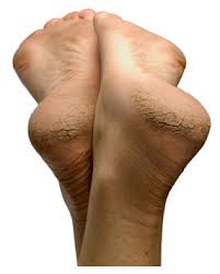
The subtalar joint, also known as the talocalcaneal joint, is one of the 33 joints in the foot. It is located at the meeting point of the talus bone and the calcaneus (heel bone), below the ankle joint. It is this joint that can cause the foot and ankle to ‘roll in’ giving the appearance of flat feet, called ‘over-pronation’.
The subtalar joint movement and range of motion is transferred up into the leg by rotating the tibia bone and also forward into the foot, controlling the locking and unlocking of another foot joint called the midtarsal joint, all necessary for normal standing, walking and running.
Over-pronation at the ankle can cause excess strain and pain to the knee
Over-pronation of the Subtalar Joint & the Knee
The effect of the subtalar joint on the knee as it causes the internal and external rotation of the tibia is a change in the alignment of the knee patella and the patella tendon. When the subtalar joint is misaligned, this rotation of the tibia causes the patella to ride abnormally with the articulation with the femur in the thigh.
This is a common root cause of a knee condition called chrondomalacia patella, advises Chief Podiatrist Michelle Champlin. Additionally, with excessive rotation of the tibia, the collateral knee ligaments are strained. Left untreated, this can result in arthroscopic knee surgeries to correct the damage caused ultimately by a misaligned subtalar joint in the foot. Even after knee surgery, unless the subtalar joint over-pronation is addressed, the knee joint degeneration will continue.
Other issues caused by over-pronation of the subtalar joint include ‘acquired’ flat feet. This is a biomechanics issue that was not present at birth and gets worse over time. However, this can generally be corrected. Travelling up the body, joints at the ankles, knees and hips can be affected. The over-pronating subtalar joint can also cause problems in the smaller joints of the forefoot and toes, leading to bunions, bunionettes, neuromas and hammer/claw toes.
Diagnosis
The most accurate diagnosis of subtalar joint over-pronation is made by a skilled Podiatrist observing the patient walking, standing and non weight bearing, as well as performing a hands on evaluation and manipulation of the foot and ankle. Observation of the foot in a walking examination is most reliable. All together, this is called a lower limb biomechanical assessment.
Treatment
Over-pronation is best treated early. There is no recommended home treatment other than the general avoidance of prolonged weight bearing in non-supportive footwear such as flip flops or ballet pumps until the patient can be assessed and treated by a Podiatrist.
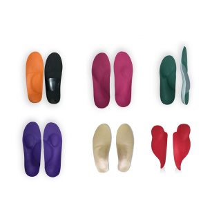
Treatment with custom-molded foot orthotics worn in regular footwear or sports shoes is critical to maintain stability of the foot and ankle and to improve the foot posture. The purpose of the orthotics is to get the subtalar joint functioning properly on its own again by tightening and toning the supporting ligaments, tendons and muscles.
As with all progressive medical conditions, it is best to start treatment early. Chief Podiatrist Michelle Champlin of Dubai Podiatry Centre recommends that all school age children are screened at age 4-5 initially for this reason.
Orthotics
Custom orthotic treatment directed at alleviating symptoms such as arch, knee or hip pain and slowing or halting the progression is the goal.
The custom orthotics must be made to the unique specifications of the patient if they are to achieve the desired results. They must also be constructed to fit in regular footwear, with Dubai Podiatry Centre’s orthotics being on average only several millimeters thick, with a variety of custom designs to accommodate even slim fitting dress shoes, football cleats or regular running shoes. It is important to the experts at Dubai Podiatry Centre that the orthotics are fitted properly into the patient’s footwear, which is one of the reasons they see the patients with their footwear and fit the orthotics into it, having the patient try the orthotics on and walk in them.
To capture the correct prescription of the orthotics, Podiatrists must have a through understanding of lower leg function. At Dubai Podiatry Centre, the Podiatrists take a specific mold of the patient’s feet, in a specific position and based upon their measurements of the subtalar joints and midtarsal joints of the foot and their ranges of motion. The Podiatrists take these measurements and then evaluate them relative to a weight bearing gait analysis.
Thorough biomechanical assessment takes place in the Dubai clinic on Sheikh Zayed Road, where the Podiatrist has access to their detailed instruments, materials and equipment.
To improve results for the patient further, the Podiatrists at Dubai Podiatry Centre have access to their own in-clinic orthotic laboratory. The Podiatrists understand the proper construction of a corrective orthotic and hand make their own patients’ orthotics, ensuring complete control over the patient’s treatment and the highest quality orthotic.
A critical element in the successful treatment of knee and foot issues with custom corrective orthotics is for the treating Podiatrist, who has the training, knowledge and experience in the treatment of these issues, to maintain total control of the casting, measuring and fabrication of the orthotics.
Health Insurance & Orthotics
Occasionally, should a patient’s the health insurance plan set their fees below a level to reimburse the patient for their Podiatrist’s time, expertise and laboratory costs, then the patient may delay treatment or seek alternative insoles or providers not equipped to meet the patients needs, which in time usually results in the problem not being properly corrected, further degeneration of the knee or other affected joints and ultimately a more costly financial outcome. Dubai Podiatry Centre’s Podiatrists provide all patients with a detailed medical report outlining expected costs for patients to send to their Insurer to seek pre-authorisation and to understand the extent of their reimbursement and coverage.
If you are concerned that your feet appear ‘flat’ or have associated knee, ankle or hip pain see a specialist biomechanics Podiatrist, who has an in-house clinic to make corrective orthotics and provide important follow up monitoring to change the prescription and monitor improvement. Consultation with the expert Podiatrists at Dubai Podiatry Centre can be made on +971 4 3435390.
The navicular bone in humans is one of the tarsal bones, found in the foot. Its name comes from the bone’s resemblance to a small boat, caused by its very concave proximal articular surface.
The navicular bone is located on the medial (upper inside) side of the foot, and connects the talus (ankle bone) behind it, with the three cuneiform bones in front of it, and to the side with the cuboid. The navicular bone forms a very important joint – the talocalcaneonavicular joint – with the talus (ankle bone) and calcaneum (heel bone). This joint transmits the entire weight of the body through the foot.
The navicular bone, in orange, sits with the cuneiform bones in front of it and talus bone behind it
It is the last of the foot’s 26 bones to start ossification (laying down bone) and does not tend to do so until the end of the second year in girls and the beginning of the fourth year in boys, although this can vary from person to person.
The tibialis posterior is the only muscle that attaches to the navicular bone.
Navicular Injuries
Injuring the navicular bone is relatively rare. Fracture of the navicular though can occur usually in athletes whilst kicking, sprinting, twisting, or falling. This will cause pain and limping. Treatment options for injury to the navicular include utilizing a non-weight bearing aircast to hold the bone stable so it can mend. If surgery is recommended, screws will be placed into the bone to help secure it. The patient will wear a cast after the surgery. In many cases, pain associated with injuries to the bone will lessen, and later return when that part of the foot is manipulated. As a result, the Podiatrist or Surgeon will manually check the navicular following treatment to see if there is pain when the top of the foot is manipulated.
The other problem that can affect the navicular bone is ‘accessory navicular syndrome’. Occasionally, some people are born with an extra bone or piece of cartilage, just above the arch on the inside of the foot. This may cause no symptoms and be entirely harmless. In some cases, the bone or the posterior tibial tendon attaching to the navicular becomes inflamed and painful. This can be caused by a trauma such as an ankle sprain, footwear rubbing on this bone or being extremely active. Many people with this extra bone will also have flat feet, putting more strain on the tendon in this area also. This syndrome tends to develop in teenage years.
Treatment
Following a thorough biomechanical assessment and medical history, the Podiatrist may recommend a range of treatments such as immobilization with an aircast, icing, NSAIDs for pain and inflammation, or custom orthotics to raise the arch and correct foot posture. If conservative treatment options do not provide relief,
If you or your child experience pain, swelling or irritation of your foot arch, contact the foot and ankle specialist clinic, Dubai Podiatry Centre on +971 4 3435390 for rapid diagnosis and treatment.
بقلم ميشيل شامبلين
الإفراط في الميلان وركبتيك
المفصل تحت الكاحل، المعروف أيضًا باسم المفصل الكاحلي العقبي، هو واحد من 33 مفصلًا في القدم. وهو يقع عند نقطة التقاء عظم الكاحل والعقب (عظم الكعب)، أسفل مفصل الكاحل. هذا المفصل هو الذي يمكن أن يتسبب في “تدحرج” القدم والكاحل مما يعطي مظهر القدم المسطحة، وهو ما يسمى “الميلان الزائد“.
يتم نقل حركة المفصل تحت الكاحل ونطاق الحركة إلى الأعلى إلى الساق عن طريق تدوير عظم الظنبوب وإلى الأمام أيضًا في القدم، للتحكم في قفل وفتح مفصل قدم آخر يسمى المفصل الوسطي، وكلها ضرورية للوقوف والمشي والجري بشكل طبيعي.
الإفراط في الميلان في الكاحل يمكن أن يسبب الضغط الزائد والألم في الركبة
الإفراط في الميلان للمفصل تحت الكاحل والركبة
تأثير المفصل تحت الكاحل على الركبة حيث أنه يسبب الدوران الداخلي والخارجي للظنبوب هو تغيير في محاذاة رضفة الركبة ووتر الرضفة. عندما يكون المفصل تحت الكاحل منحرفًا، يؤدي هذا الدوران للظنبوب إلى تحرك الرضفة بشكل غير طبيعي مع المفصل مع عظم الفخذ في الفخذ.
هذا هو السبب الجذري الشائع لحالة الركبة التي تسمى تلين الرضفة، كما تنصح رئيسة أطباء الأقدام ميشيل شامبلين. بالإضافة إلى ذلك، مع الدوران المفرط لعظم الساق، تتوتر الأربطة الجانبية للركبة. إذا تُرك هذا الأمر دون علاج، فقد يؤدي ذلك إلى إجراء عمليات جراحية بالمنظار في الركبة لتصحيح الضرر الناتج في النهاية عن محاذاة المفصل تحت الكاحل بشكل غير صحيح في القدم. حتى بعد جراحة الركبة، ما لم تتم معالجة فرط كب المفصل تحت الكاحل، فإن انحطاط مفصل الركبة سيستمر
تشمل المشكلات الأخرى الناجمة عن الإفراط في كب المفصل تحت الكاحل الأقدام المسطحة “المكتسبة”. هذه مشكلة في الميكانيكا الحيوية لم تكن موجودة عند الولادة وتزداد سوءًا بمرور الوقت. ومع ذلك، يمكن تصحيح هذا بشكل عام. أثناء السفر إلى أعلى الجسم، يمكن أن تتأثر المفاصل عند الكاحلين والركبتين والوركين. يمكن أيضًا أن يسبب المفصل تحت الكاحل المفرط النطق مشاكل في المفاصل الأصغر في مقدمة القدم وأصابع القدم، مما يؤدي إلى الأورام والأورام العصبية وأصابع القدم المطرقية/المخلبية.
تشخبص
يتم إجراء التشخيص الأكثر دقة للكب الزائد للمفصل تحت الكاحل من قبل طبيب أقدام ماهر يراقب المريض أثناء المشي والوقوف وعدم تحمل الوزن، بالإضافة إلى إجراء تقييم عملي والتلاعب بالقدم والكاحل. تعتبر مراقبة القدم أثناء فحص المشي هي الأكثر موثوقية. يُطلق على هذا معًا تقييم الميكانيكا الحيوية للطرف السفلي.
علاج
من الأفضل علاج الإفراط في الكب مبكرًا. لا يوجد علاج منزلي موصى به بخلاف التجنب العام لتحمل الوزن لفترة طويلة بارتداء أحذية غير داعمة مثل الصنادل أو أحذية الباليه حتى يمكن تقييم المريض وعلاجه بواسطة طبيب الأقدام.
يعد العلاج باستخدام أجهزة تقويم القدم المصممة خصيصًا والتي يتم ارتداؤها في الأحذية العادية أو الأحذية الرياضية أمرًا بالغ الأهمية للحفاظ على ثبات القدم والكاحل وتحسين وضعية القدم. الغرض من تقويم العظام هو جعل المفصل تحت الكاحل يعمل بشكل صحيح من تلقاء نفسه مرة أخرى عن طريق شد وتنعيم الأربطة والأوتار والعضلات الداعمة.
كما هو الحال مع جميع الحالات الطبية المتقدمة، فمن الأفضل أن تبدأ العلاج في وقت مبكر. توصي رئيسة أطباء الأقدام ميشيل شامبلين من مركز دبي لعلاج الأرجل بإجراء فحص لجميع الأطفال في سن المدرسة في سن 4-5 سنوات في البداية لهذا السبب
تقويم العظام
الهدف هو العلاج التقويمي المخصص الذي يهدف إلى تخفيف الأعراض مثل آلام القوس أو الركبة أو الورك وإبطاء أو إيقاف التقدم.
يجب أن يتم تصنيع أجهزة تقويم العظام المخصصة وفقًا للمواصفات الفريدة للمريض إذا أراد تحقيق النتائج المرجوة. ويجب أيضًا تصميمها لتناسب الأحذية العادية، حيث يبلغ متوسط سمك أجهزة تقويم العظام في مركز دبي لطب الأقدام عدة ملليمترات فقط، مع مجموعة متنوعة من التصميمات المخصصة لاستيعاب حتى الأحذية الرسمية الضيقة، أو أحذية كرة القدم، أو أحذية الجري العادية. من المهم للخبراء في مركز دبي لطب الأرجل أن يتم تركيب أجهزة تقويم العظام بشكل صحيح في أحذية المريض، وهذا أحد الأسباب التي تجعلهم يرون المرضى بأحذيتهم ويضعون أجهزة تقويم العظام فيها، مما يجعل المريض يجرب جهاز تقويم العظام ويمشي. فيهم.
للحصول على الوصفة الطبية الصحيحة لتقويم العظام، يجب أن يكون لدى أطباء الأقدام فهم شامل لوظيفة الجزء السفلي من الساق. في مركز دبي لعلاج الأرجل، يأخذ أطباء الأرجل قالبًا محددًا لقدم المريض، في وضع محدد وبناءً على قياساتهم للمفاصل تحت الكاحل والمفاصل الوسطى للقدم ونطاقات حركتها. يأخذ أطباء الأقدام هذه القياسات ثم يقومون بتقييمها مقارنة بتحليل المشية الذي يحمل الوزن.
يتم إجراء تقييم ميكانيكي حيوي شامل في عيادة دبي الواقعة على طريق الشيخ زايد، حيث يمكن لطبيب الأقدام الوصول إلى أدواته ومواده ومعداته التفصيلية.
لتحسين النتائج للمريض بشكل أكبر، يتمتع أطباء الأقدام في مركز دبي لعلاج الأرجل بإمكانية الوصول إلى مختبر تقويم العظام الخاص بهم داخل العيادة. يفهم أطباء الأقدام البناء الصحيح لجهاز تقويم العظام التصحيحي ويقومون بتصنيع أجهزة تقويم العظام لمرضاهم يدويًا، مما يضمن التحكم الكامل في علاج المريض وتقويم العظام بأعلى جودة.
أحد العناصر الحاسمة في العلاج الناجح لمشاكل الركبة والقدم باستخدام تقويم العظام التصحيحي المخصص هو أن يقوم طبيب الأقدام المعالج، الذي لديه التدريب والمعرفة والخبرة في علاج هذه المشكلات، بالحفاظ على التحكم الكامل في صب وقياس وتصنيع القدم. تقويم العظام
التأمين الصحي وتقويم العظام في بعض الأحيان، إذا حددت خطة التأمين الصحي للمريض رسومه أقل من المستوى لتعويض المريض عن وقت طبيب الأقدام وخبرته وتكاليف المختبر، فقد يؤخر المريض العلاج أو يبحث عن نعال بديلة أو مقدمي خدمات غير مجهزين لتلبية هذه المتطلبات. احتياجات المرضى، والتي عادة ما تؤدي مع مرور الوقت إلى عدم تصحيح المشكلة بشكل صحيح، وزيادة تدهور الركبة أو المفاصل المتضررة الأخرى، وفي النهاية نتيجة مالية أكثر تكلفة. يقدم أطباء الأقدام في مركز دبي لعلاج الأقدام لجميع المرضى تقريرًا طبيًا مفصلاً يوضح التكاليف المتوقعة للمرضى لإرساله إلى شركة التأمين الخاصة بهم للحصول على إذن مسبق وفهم مدى تعويضهم وتغطيتهم.
إذا كنت قلقًا من أن تبدو قدميك “مسطحة” أو تعاني من آلام في الركبة أو الكاحل أو الورك، فراجع أخصائي طب الأقدام المتخصص في الميكانيكا الحيوية، والذي لديه عيادة داخلية لصنع تقويم العظام التصحيحية وتوفير مراقبة متابعة مهمة لتغيير الوصفة الطبية ومراقبة التحسن. . يمكن إجراء الاستشارة مع خبراء طب الأقدام في مركز دبي لعلاج الأقدام على الرقم +971 4 3435390.
العظم الزورقي عند الإنسان هو أحد العظام الرصغية الموجودة في القدم. يأتي اسمها من تشابه العظم مع قارب صغير، بسبب سطحه المفصلي القريب المقعر للغاية.
يقع العظم الزورقي على الجانب الإنسي (العلوي من الداخل) من القدم، ويربط الكاحل (عظم الكاحل) خلفه، مع العظام المسمارية الثلاثة الموجودة أمامه، وإلى الجانب مع المكعب. يشكل العظم الزورقي مفصلًا مهمًا جدًا – المفصل الكاحلي الزورقي الزورقي – مع الكاحل (عظم الكاحل) والعقبي (عظم الكعب). ينقل هذا المفصل وزن الجسم بالكامل عبر القدم.
يقع العظم الزورقي، باللون البرتقالي، والعظام المسمارية أمامه وعظم الكاحل خلفه
وهي آخر عظمة من عظام القدم البالغ عددها ٢٦ عظمة تبدأ بالتعظم (وضع العظم) ولا تميل إلى ذلك حتى نهاية السنة الثانية عند البنات وبداية السنة الرابعة عند الأولاد، على الرغم من أن هذا يمكن أن يختلف من شخص إلى آخر. شخص.
الظنبوب الخلفي هو العضلة الوحيدة التي ترتبط بالعظم الزورقي
إصابات البحرية
من النادر نسبياً إصابة العظم الزورقي. على الرغم من ذلك، يمكن أن يحدث كسر في الزورقي عادةً عند الرياضيين أثناء الركل أو الركض أو الالتواء أو السقوط. هذا سوف يسبب الألم والعرج. تشمل خيارات العلاج لإصابة الزورقي استخدام جهاز هوائي لا يحمل وزنًا للحفاظ على استقرار العظام حتى تتمكن من الإصلاح. إذا أوصى بإجراء عملية جراحية، فسيتم وضع براغي في العظم للمساعدة في تثبيته. سيرتدي المريض جبيرة بعد الجراحة. في كثير من الحالات، يقل الألم المرتبط بإصابات العظام، ويعود لاحقًا عند التلاعب بهذا الجزء من القدم. ونتيجة لذلك، سيقوم طبيب الأقدام أو الجراح بفحص المنطقة الزورقية يدويًا بعد العلاج لمعرفة ما إذا كان هناك ألم عند التلاعب بأعلى القدم.
المشكلة الأخرى التي يمكن أن تؤثر على العظم الزورقي هي “المتلازمة الزورقية الإضافية”. في بعض الأحيان، يولد بعض الأشخاص بعظم إضافي أو قطعة من الغضروف، أعلى القوس مباشرة من داخل القدم. قد لا يسبب هذا أي أعراض ويكون غير ضار تمامًا. في بعض الحالات، يصبح العظم أو الوتر الظنبوبي الخلفي المتصل بالزورقي ملتهبًا ومؤلمًا. يمكن أن يحدث هذا بسبب صدمة مثل التواء الكاحل أو احتكاك الأحذية بهذا العظم أو النشاط الشديد. العديد من الأشخاص الذين لديهم هذا العظم الزائد سيكون لديهم أيضًا أقدام مسطحة، مما يزيد الضغط على الوتر في هذه المنطقة أيضًا. تميل هذه المتلازمة إلى التطور في سنوات المراهقة.
العلاج: بعد إجراء تقييم ميكانيكي حيوي شامل والتاريخ الطبي، قد يوصي طبيب الأقدام بمجموعة من العلاجات مثل التثبيت باستخدام الهواء، أو وضع الثلج، أو مضادات الالتهاب غير الستيروئيدية للألم والالتهاب، أو تقويم العظام المخصص لرفع القوس وتصحيح وضعية القدم. إذا لم توفر خيارات العلاج التحفظي الراحة،
إذا كنت تعاني أنت أو طفلك من ألم أو تورم أو تهيج في قوس قدمك، اتصل بالعيادة التخصصية للقدم والكاحل في مركز دبي للعناية بالاقدام على الرقم ٠٤٣٤٣٥٣٩٠ للتشخيص والعلاج السريع.


 Written by Michelle Champlin BSc Pod., M.Ch.S., S.R., Ch., (UK)