
fbpx

Please wait...


BLOG

No more posts

One of the most common questions we hear at Dubai Podiatry Centre is: “Why won’t my nail fungus clear, even though I’ve tried home remedies?”

The answer is that not all fungal nail infections are the same.

Different types of nail fungus affect different parts of the toenail, and each type requires a different treatment approach. This is why a treatment that works for one person may have little or no effect for another.


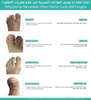
Why home remedies often fail to cure nail fungus showing fungal infections in the nail root, under the nail, superficial nail fungus, and nail plate with appropriate treatment options

Superficial White Nail Fungus

One of the easiest types of fungal infection to treat is superficial white fungal infection.

This commonly develops in warm, humid conditions inside closed footwear and under nail polish. The fungus affects only the surface of the nail and often appears as white, chalky or powdery patches on the top of the toenail.

Because the infection is only on the surface, the affected layer can often be gently reduced with professional nail filing or careful buffing, allowing a topical antifungal nail lacquer to reach the remaining fungus more effectively.

As the nail gradually grows out, the healthy nail replaces the infected portion.


Fungus Growing Beneath the Nail

A more difficult type of fungal infection develops underneath the nail plate.

This is commonly associated with a widespread fungal skin infection affecting the sole of the foot, often referred to as moccasin-type athlete’s foot. The fungus can spread beneath the toenail, where it becomes protected by the thick nail plate.

When the fungus is trapped underneath the nail, simply painting antifungal medication onto the surface often has limited success because the medication cannot adequately reach the infection.


Depending on the severity of the infection, treatment may involve oral antifungal medication, careful reduction of the thickened nail, or, in selected cases, nail avulsion of the toenail to allow direct treatment of the underlying nail bed as the new nail grows.

Following nail removal, the smaller toenails generally regrow in approximately three months, while the big toenail may take anywhere from six months to one year to fully regrow.


Fungus That Reaches the Nail Root

In some patients, the fungal infection extends into the nail root (matrix), where the nail is produced.

When this occurs, oral antifungal medication is often recommended because the medication reaches the growing nail through the bloodstream, allowing the new nail to grow free of infection.

If there is also extensive fungal material trapped beneath a thick or damaged nail plate, reducing or removing the affected nail may be recommended to improve access for topical treatment and encourage healthy nail regrowth.


When Oral Medication Is Not Suitable

Before prescribing oral antifungal medication, liver function blood tests are often performed to ensure the medication can be taken safely.

Occasionally, these tests identify previously undiagnosed liver abnormalities. In these situations, even patients who feel completely healthy may not be suitable candidates for oral antifungal medication.

For these patients, treatment may focus on physical methods, including removal of the infected toenail where appropriate, combined with topical antifungal medication applied directly to the exposed nail bed after healing. This allows the new nail to grow in an environment with a much lower fungal burden.


Why Professional Assessment Matters

Many people spend months or even years trying home remedies without success, simply because they are treating a different type of fungal infection.

Successful treatment begins with identifying exactly where the fungus is located, whether it is on the surface of the nail, beneath the nail plate, or involving the nail root.

Once the type and stage of the infection have been identified, the most appropriate treatment plan can be chosen, giving the best opportunity for healthy, fungus-free nail growth.


For more information or to book an appointment please call our clinic +971 4 3435390 or WhatsApp +971 50 3553024


One of the most common questions patients ask at Dubai Podiatry Centre is:

“Am I too old to treat my flat feet?”

The simple answer is no.

Many people believe that flat feet can only be treated during childhood and that once you become an adult, nothing more can be done. Fortunately, this is one of the biggest misconceptions we hear in clinic.

Not Everyone Diagnosed with Flat Feet Actually Has Flat Feet

There are several different types of flat feet, but the most common condition we see is not a true flat foot at all.

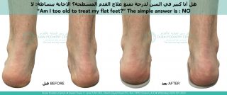
In many cases, the ankle leans inwards, causing the arch to collapse under body weight. The arch then appears flat, even though the foot itself still has the ability to regain its natural shape once the ankle is correctly aligned.

Rather than thinking of the foot as permanently flat, it is often more accurate to think of it as an arch that has collapsed because the ankle is no longer supporting it correctly.


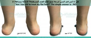
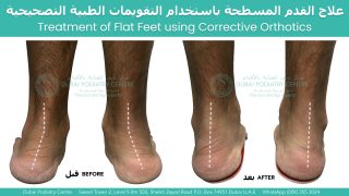
Adult flat feet before and after custom orthotics showing improved heel alignment and foot posture

Child with flat feet before and after podiatry treatment using custom orthotics showing corrected heel alignment


Adult Treatment Can Actually Be Faster

Many people are surprised when we explain that correcting this type of flat foot can actually progress faster in adults than in children.

Children have high levels of human growth hormone, which keeps their ligaments soft, flexible and easy to manipulate. This makes repositioning the bones relatively straightforward and usually very comfortable.

However, because the ligaments are constantly growing and remodelling, they often take longer to adapt and stabilise in their new position. For this reason, children commonly progress through each stage of treatment over approximately three to six months.

Adults are different.

Adult ligaments are firmer and less elastic, so there can be slightly more resistance when repositioning the foot. For this reason, we usually make smaller adjustments at each stage.

Once the bones have been repositioned, however, the adult foot often adapts surprisingly quickly. Instead of waiting three to six months between stages, many adults can progress every one to three months, allowing treatment to advance more rapidly.

Similar to Orthodontic Braces

A useful comparison is orthodontic braces for the teeth.

Most people understand that braces are not just for children. Adults successfully wear braces every day to gradually reposition their teeth into better alignment.

Corrective orthotics work on a similar principle.

Instead of moving teeth, they gently reposition the bones of the feet and ankles in carefully planned stages. As the alignment improves, the arch is supported more effectively and, in suitable cases, the foot can gradually regain a healthier arch shape.

Each new stage builds upon the previous one, allowing gradual correction rather than trying to make one large adjustment all at once.


It’s Never Too Late to Have an Assessment

If you have been told you have flat feet, it does not necessarily mean your feet are permanently flat.

The first step is determining why your arch has collapsed. For many patients, the underlying issue is ankle alignment rather than the shape of the foot itself.

With the correct diagnosis and a staged treatment plan, adults can often achieve significant improvements in foot alignment, comfort and function.

So if you’ve been wondering whether you’ve left it too late to treat your flat feet, the answer may be much more encouraging than you expected.

At Dubai Podiatry Centre, we regularly assess both children and adults and develop personalised treatment plans based on the specific cause of each patient’s foot alignment, because no two cases of flat feet are exactly the same.


For more information or to book an appointment please call our clinic +971 4 3435390 or WhatsApp +971 50 3553024


Plantar fasciitis is one of the most common causes of heel pain. People typically notice it when they take their first steps out of bed in the morning or when they stand after sitting for a prolonged period. The pain often eases after walking for a few minutes, only to return again after periods of rest.


What is the plantar fascia?

The plantar fascia is a strong band of connective tissue that runs from the heel bone (calcaneus) to the base of the toes. Its primary function is to help support the arch of the foot.

When you walk, run or land from a jump, the plantar fascia becomes tensioned, helping the arch maintain its shape and absorb forces. Without this support, the bones of the midfoot would be subjected to much greater mechanical stress.

A healthy plantar fascia is not designed to remain under constant tension. Instead, it should be relatively relaxed at rest and become taut only when required during movement and weight-bearing activities.


Why does plantar fasciitis develop?

The term plantar fasciitis literally means inflammation associated with the plantar fascia. Although the pain is felt in the heel, the discomfort is usually concentrated where the plantar fascia attaches to the heel bone.

Rather than viewing plantar fasciitis as simply an isolated heel problem, it is often more helpful to consider why the plantar fascia has become overloaded in the first place.

In many people, the ligaments and muscles of the foot provide enough stability to support the arch without excessive strain on the plantar fascia. However, if the alignment of the foot changes, the plantar fascia may remain under tension for much longer than it was designed to.

One of the most common contributing factors is excessive pronation of the foot and ankle. As the foot rolls inward, the plantar fascia may become progressively tensioned throughout standing and walking instead of only during the phases when it is normally required.

An easy way to understand the problem

Imagine holding a single strand of long hair with a heavy weight attached to the end.

The hair itself is not painful.

The pain would occur where the hair is anchored into the scalp because that attachment point is constantly being pulled.

Once the weight is removed, the strain at the attachment point disappears.

The plantar fascia behaves in a similar way. The discomfort usually develops where the fascia attaches into the heel bone rather than within the fascia itself. If excessive tension continues day after day, this attachment site can become irritated and painful.


Why is the pain worse in the morning?

Many people notice that the first few steps in the morning are the most painful.

After sleeping or sitting for a prolonged period, blood flow and tissue fluid movement slow down. When the foot is suddenly loaded, the already irritated attachment of the plantar fascia is placed under tension again, producing the characteristic sharp heel pain.

As you begin walking, the calf muscles act as a pump, improving circulation through the lower leg and foot. This often reduces discomfort temporarily, which explains why many people feel better after several minutes of walking.

Treating the cause—not just the symptoms

While heel cushions, supportive insoles and stretching programmes may provide symptom relief for some individuals, long-term improvement depends on identifying and correcting the mechanical cause of the overload.

If excessive pronation or another alignment abnormality is placing unnecessary tension on the plantar fascia, simply adding cushioning beneath the heel may not adequately address the underlying problem.

A corrective orthotic is designed to improve foot and ankle alignment rather than simply support the arch. Because every person walks differently, the correction required for the left foot is often different from that required for the right foot. The amount of correction should therefore be individually prescribed.

As the abnormal strain on the plantar fascia is reduced over time, the attachment at the heel is given the opportunity to recover, allowing symptoms to gradually settle while also addressing the underlying biomechanical problem that contributed to the condition.

The key message

Plantar fasciitis should not simply be viewed as inflammation of the heel. In many cases, it is the result of an underlying mechanical imbalance that places excessive and prolonged tension on the plantar fascia where it attaches to the heel bone.

Successful long-term treatment focuses on identifying the biomechanical cause, correcting the alignment of the foot and ankle, and reducing the abnormal forces responsible for the condition. By addressing the underlying mechanics rather than simply cushioning the symptoms, many patients can achieve lasting relief and reduce the likelihood of recurrence.


For more information or to book an appointment please call our clinic +971 4 3435390 or WhatsApp +971 50 3553024


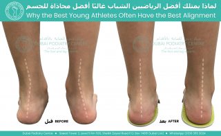
Before and after foot alignment correction in a young athlete showing reduced pronation, improved heel position, and enhanced lower limb biomechanics with corrective orthotics


When we watch elite athletes perform, we often admire their speed, strength, agility, and coordination. What many people do not realise is that beneath these athletic abilities lies something equally important: good biomechanical alignment.

In many cases, the young athletes who successfully progress through junior sport and eventually reach professional levels are those whose bodies remain symmetrical, balanced, and resistant to repetitive injury.


The Importance of Lower Limb Alignment

The feet are the foundation of the body’s kinetic chain. Every step, jump, sprint, and change of direction begins with the way the feet interact with the ground.

When the subtalar joints of the feet and ankles become excessively pronated (rolling inward), a chain reaction can occur throughout the lower limb:

    • Internal rotation of the tibia (shin bone)
    • Increased stress on the knee joint
    • Altered hip mechanics
    • Anterior pelvic tilt
    • Reduced postural efficiency
    • Increased muscular compensation throughout the body

As a result, running mechanics become less efficient and athletic performance can suffer.


Why Some Young Athletes Fall Behind

It is common to see children who are naturally talented at a young age begin to struggle as sporting demands increase.

Around the ages of 10–16 years, training intensity often increases dramatically. During this period, poor lower limb alignment may begin to reveal itself through repetitive injuries rather than obvious pain at first.

Common problems may include:

    • Medial knee pain
    • Recurrent groin strains
    • Patellofemoral pain
    • Achilles tendon discomfort
    • Shin splints
    • Ankle instability
    • Recurrent muscle tightness
    • Increased fatigue during training

A football player may develop repeated groin strains and knee pain when changing direction. A rugby player may suffer ongoing knee injuries during contact and sprinting activities. A tennis player may experience instability while lunging, accelerating, or moving backwards to retrieve shots.

Unfortunately, many promising young athletes spend more time recovering from injuries than participating in training. By the mid-teenage years, some are unable to continue progressing through elite development pathways because their bodies simply cannot tolerate the physical demands being placed upon them.


The Role of Modern Biomechanical Assessment

Advances in biomechanical assessment technology now allow clinicians to identify alignment issues far earlier than ever before.

By analysing posture, gait, foot function, lower limb alignment, and movement patterns, it is often possible to identify the underlying mechanical stresses that contribute to repetitive injuries.

In many cases, excessive pronation of the subtalar joint is a significant contributing factor.

When the foundation is unstable, the structures above it must compensate.

Corrective Orthotics vs Accommodative Orthotics

Not all orthotics are designed with the same purpose.

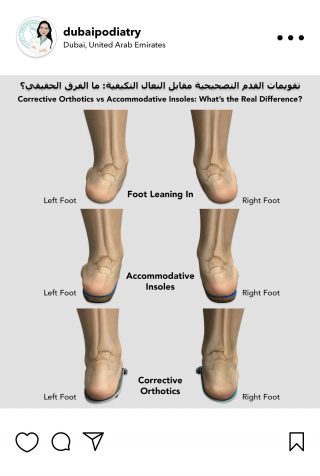
Comparison of foot alignment without support, with accommodative insoles, and with corrective orthotics, demonstrating the effect of orthotic devices on foot posture and heel alignment


Accommodative orthotics are primarily designed to cushion and support the foot. While they may provide comfort, they do not necessarily correct abnormal alignment.

Corrective orthotics are designed to influence foot and ankle position and improve lower limb biomechanics. By improving subtalar joint alignment, corrective orthotics may help:

    • Improve foot stability
    • Reduce excessive internal rotation
    • Improve knee alignment
    • Improve pelvic positioning
    • Reduce abnormal stress on muscles and ligaments
    • Reduce the risk of repetitive overuse injuries

For growing athletes, maintaining good alignment throughout adolescence can be particularly important because the body is continually developing and adapting.


Investing in an Athlete’s Future

Every young athlete dreams of reaching their full potential.

Talent, commitment, coaching, and hard work are all essential ingredients for success. However, maintaining a body that can tolerate years of training is equally important.

A comprehensive biomechanical assessment can help identify whether poor lower limb alignment is placing a young athlete at increased risk of injury. Early intervention may help reduce repetitive strain, improve movement efficiency, and keep athletes participating in the sport they love.

For many aspiring footballers, rugby players, tennis players, runners, and other athletes, addressing alignment early may be one of the most important investments they can make in their future sporting career.

At Dubai Podiatry Centre, we believe that injury prevention starts with proper alignment. The earlier biomechanical issues are identified, the greater the opportunity to support healthy growth, efficient movement, and long-term athletic development.


For more information or to book an appointment please call our clinic +971 4 3435390 or WhatsApp +971 50 3553024


A patient attended our clinic today complaining of severe pain underneath the outside of his left foot. The pain had become so intense that he was limping and described the sensation as walking on a sharp piece of glass.

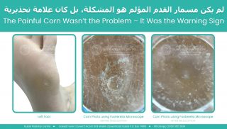
On examination, we found a hard corn underneath the fifth metatarsal head on the outside border of his left foot. Many people assume that a corn is the problem, but in reality a corn is often the body’s attempt to protect itself from excessive pressure.

We removed the corn painlessly, and the patient experienced immediate relief. However, we explained that unless we addressed the underlying cause, the corn would simply return.

To investigate further, we performed a 3D body scan and biomechanical assessment.

The results were fascinating.

The patient had a significant imbalance throughout his body. His left leg was functionally longer than his right. His pelvis was tilted, with the right side anteriorly tilted and the left side posteriorly tilted. He was carrying considerably more body weight through the left side of his body, creating excessive pressure beneath the outside of the left foot.

His medical history helped explain the pattern. He had previously undergone two lower back surgeries on the left side, suffered left hip problems, torn ACL injuries involving the left knee, and undergone left Achilles tendon surgery.

Over many years, his body had adapted to these injuries.

The scan showed that his pelvis had shifted towards the left side, causing increased loading through the left leg and foot. At the same time, his right foot was internally rotated and functioning almost like a golfer at the top of a backswing position. This created high tensile stress through the right Achilles tendon, the right knee, and the lower back while simultaneously increasing compression forces through the left side of the body.

The painful corn underneath the left foot was simply the area where the body was absorbing the greatest amount of pressure.

The development of a corn follows a predictable sequence.

First, the body produces a layer of hard skin to protect itself. This creates a broad plaque of callus, similar to a pancake of thicker skin.

If the pressure continues, the body creates a concentrated plug of dense keratin at the centre of the pressure point. Keratin is the same material found in toenails. This hard central core forms what we recognise as a hard corn.

As pressure continues to increase, inflammation develops beneath the area. Patients often describe burning, stinging, or sharp sensations. This was the stage our patient had reached.

If left untreated, the next stage can be the formation of a soft corn. The tissues become saturated with inflammatory fluid and appear white, rubbery, and extremely painful.

The final stage is tissue breakdown and ulceration.

This progression is particularly important in patients with diabetes, neuropathy, or vascular disease. These patients may not feel the early warning signs of pressure and pain. As a result, the protective stages can progress unnoticed until an ulcer develops.

Many patients believe ulcers appear suddenly and without warning. In reality, the body often gives multiple warning signals beforehand, but if sensation is reduced, those signals may not be recognised.

For this patient, the solution was not repeated corn removal.

The solution was correcting the forces causing the corn.

We prescribed custom corrective orthotics designed specifically for his individual biomechanics. The right orthotic was intentionally very different from the left because each side of the body required a different correction.

The aim was to improve pelvic balance, reduce the functional leg length discrepancy, redistribute pressure more evenly between both feet, and decrease excessive loading through the left foot.

We performed further scans with the orthotics in place and saw immediate improvements in alignment and pressure distribution.

The body can be compared to a wobbly table. If one table leg is shorter than the others, the tabletop becomes uneven. Placing a carefully measured support beneath the shorter leg restores balance.

Corrective orthotics work in a similar way. While we cannot make one leg physically grow longer, we can improve balance and alignment so that the body functions more efficiently and with less stress.

The patient will return in three months for reassessment. As the body adapts to improved alignment, muscle tension, posture, and weight distribution can all change. Repeat scanning will allow us to monitor these changes and determine whether further orthotic adjustments are required.

The important lesson from this case is that a corn is rarely just a skin problem.

A corn is often a warning sign that excessive pressure is travelling through a specific part of the foot. Removing the corn may provide temporary relief, but identifying and correcting the underlying mechanical cause is what provides a long-term solution.


For more information or to book an appointment please call our clinic +971 4 3435390 or WhatsApp +971 50 3553024


Before and after treatment of adult flat feet using custom corrective orthotics, showing improved heel alignment and foot posture from the rear view at Dubai Podiatry Centre.

Flat feet are commonly associated with inward rolling of the ankles, a condition known as overpronation. When the ankles tilt inward, the body’s weight is distributed unevenly across the feet, which can lead to foot pain, poor posture, and increased strain on the knees, hips, and lower back.

Custom corrective orthotics are designed to address the root cause of the problem by improving foot and ankle alignment. Rather than simply raising the arch, the primary objective is to reposition the heel and ankle into a more neutral and balanced position. Once proper alignment is restored, the arch often begins to improve naturally as the foot functions more efficiently.

Many people assume that treating flat feet means forcing the arch upward. However, excessively elevating the arch without correcting the underlying ankle alignment can make orthotics uncomfortable and difficult to wear. Effective orthotic therapy focuses on stabilising the heel, controlling overpronation, and supporting the foot in its optimal position.

A properly prescribed custom orthotic provides both arch support and heel correction, creating a stable foundation for standing, walking, and everyday activities. Improved alignment can enhance comfort, reduce strain on the lower limbs, and promote healthier movement patterns.

The photographs above demonstrate the effect of corrective orthotics on foot posture. By restoring proper heel and ankle alignment, the feet become more balanced, allowing for improved arch function and overall biomechanical efficiency.


For more information or to book an appointment please call our clinic +971 4 3435390 or WhatsApp +971 50 3553024


Illustration of a plantar wart on the sole of a foot, showing inward growth beneath the skin and a close-up with small black dots (blood vessels). Text highlights that it is a viral infection, may bleed if cut, and is painful with pressure.

A plantar wart, also known as a verruca, is a viral infection that occurs on the sole of the foot. Unlike warts elsewhere on the body that grow outward as raised bumps, plantar warts grow inward due to the constant pressure of standing and walking. This means they often appear flat on the surface while developing a bulb-like structure deeper within the skin.

Because of this inward growth, plantar warts can be more difficult to treat. Treatments that work well for outward-growing warts on the hands or other parts of the body, such as cryotherapy (liquid nitrogen) or salicylic acid, often have lower success rates on the sole of the foot. This is because the wart is not easily accessible on the surface, making it harder for these treatments to reach the infected tissue effectively.

الثؤلول الأخمصي، المعروف أيضًا باسم الفيروكا (Verruca)، هو عدوى فيروسية تصيب باطن القدم. وعلى عكس الثآليل التي تظهر في مناطق أخرى من الجسم وتنمو إلى الخارج على شكل نتوءات بارزة، فإن الثآليل الأخمصية تنمو إلى الداخل بسبب الضغط المستمر الناتج عن الوقوف والمشي. ولهذا السبب، غالبًا ما تبدو مسطحة على سطح الجلد، بينما تمتد في العمق على شكل كتلة تشبه البصلة داخل الأنسجة.

وبسبب هذا النمو الداخلي، تكون الثآليل الأخمصية أكثر صعوبة في العلاج. فالعلاجات التي قد تكون فعّالة للثآليل التي تنمو إلى الخارج على اليدين أو في أجزاء أخرى من الجسم، مثل العلاج بالتبريد (النيتروجين السائل) أو حمض الساليسيليك، غالبًا ما تحقق معدلات نجاح أقل عند استخدامها لعلاج الثآليل الموجودة في باطن القدم. ويعود ذلك إلى أن الثؤلول لا يكون مكشوفًا بشكل كافٍ على السطح، مما يجعل وصول هذه العلاجات إلى الأنسجة المصابة أكثر صعوبة وأقل فعالية.


So how does a plantar wart develop in the first place?

Infographic showing how plantar warts develop: infected skin cells spread in moist public areas, barefoot contact transfers the virus, it enters through small breaks in the skin, and a wart forms over months. High-risk areas listed include swimming pools, gym changing rooms, communal showers, and water parks, with related icons.The condition is caused by a virus that is spread through contact with infected skin cells. This typically occurs in high-risk environments where people walk barefoot, such as swimming pools, gym changing rooms, and water parks. An infected skin cell from another person can be shed onto the floor, and if you walk over that area within a short time frame, the virus may transfer to your foot.

However, the virus cannot easily penetrate healthy skin. The outermost layer of the skin, known as the stratum corneum, acts as a protective barrier. This layer is structured like tightly packed scales, similar to fish scales, forming a strong defence against external invasion.

When the feet are dry, this barrier remains tightly closed and highly protective. But when the skin becomes wet or sweaty for prolonged periods, the structure of this layer changes. The “scales” begin to separate slightly, creating microscopic gaps. This makes the skin more porous and vulnerable to viral entry.

At the same time, an infected foot in a moist environment can shed virus-containing skin cells more easily. These infected cells can then be picked up by another person walking over the same surface, particularly in high-traffic, damp areas.

Once the virus enters the skin, it develops slowly. It can take several months, often up to six months or more, before a plantar wart becomes visible. During this time, the virus grows within the deeper layers of the skin before gradually appearing on the surface.

فكيف يتكوّن الثؤلول الأخمصي في المقام الأول؟

تنجم هذه الحالة عن فيروس ينتقل من خلال ملامسة خلايا الجلد المصابة. ويحدث ذلك عادةً في البيئات عالية الخطورة التي يمشي فيها الأشخاص حفاة الأقدام، مثل أحواض السباحة، وغرف تبديل الملابس في الصالات الرياضية، والحدائق المائية. فقد تسقط خلية جلدية مصابة من شخصٍ ما على الأرض، وإذا مررت فوق تلك المنطقة خلال فترة قصيرة، فقد ينتقل الفيروس إلى قدمك.

ومع ذلك، لا يستطيع الفيروس اختراق الجلد السليم بسهولة. فالطبقة الخارجية من الجلد، والمعروفة باسم الطبقة القرنية، تعمل كحاجز واقٍ. وتتكوّن هذه الطبقة من خلايا متراصة بإحكام تشبه حراشف السمك، مما يشكّل خط دفاع قويًا ضد العوامل الخارجية.

عندما تكون القدمان جافتين، يبقى هذا الحاجز محكم الإغلاق ويوفر حماية عالية. ولكن عندما يصبح الجلد رطبًا أو متعرقًا لفترات طويلة، تتغير بنية هذه الطبقة. إذ تبدأ “الحراشف” الجلدية بالانفصال بشكل طفيف، مما يخلق فجوات مجهرية صغيرة. وهذا يجعل الجلد أكثر نفاذية وأكثر عرضة لدخول الفيروسات.

وفي الوقت نفسه، يمكن للقدم المصابة الموجودة في بيئة رطبة أن تطرح خلايا جلدية تحتوي على الفيروس بسهولة أكبر. ويمكن لهذه الخلايا المصابة أن تنتقل إلى شخص آخر يمشي على السطح نفسه، خاصةً في الأماكن الرطبة كثيرة الاستخدام.

وبمجرد دخول الفيروس إلى الجلد، فإنه يتطور ببطء. فقد يستغرق الأمر عدة أشهر، وغالبًا ما يصل إلى ستة أشهر أو أكثر، قبل أن يصبح الثؤلول الأخمصي مرئيًا. وخلال هذه الفترة، ينمو الفيروس داخل الطبقات العميقة من الجلد قبل أن يبدأ بالظهور تدريجيًا على السطح.

Comparison infographic of a plantar wart and a corn. The plantar wart shows small black dots (blood vessels), is a viral infection with blood supply, may bleed, and is painful with side pressure. The corn is a build-up of hard skin from pressure, has no blood supply, does not bleed, has no black dots, and is painful with direct pressure. A note explains that black dots in plantar warts are tiny blood vessels indicating active infection.Plantar warts are sometimes confused with corns, but they are very different. A corn is a build-up of hard skin caused by pressure and friction. It can often be reduced without bleeding, although it may feel like walking on a sharp point. A plantar wart, however, is a living viral lesion. If disturbed or cut, it will bleed because it has developed its own blood supply within the skin.

One of the key identifying features of a plantar wart is the presence of small black dots within the lesion. These are not roots, but tiny blood vessels known as capillaries. The virus disrupts and constricts these vessels, causing them to appear dark. Their presence is a sign that the wart is active and established within the tissue.

This is where Dermojet® treatment is particularly effective. Because plantar warts grow deep within the skin, a treatment that can reach and disrupt the infected tissue at its core is essential. Dermojet® delivers a high-pressure microjet that penetrates beneath the surface, targeting the area where the virus is thriving and improving the likelihood of successful resolution.

يتم الخلط أحيانًا بين الثآليل الأخمصية ومسامير القدم، لكنهما حالتان مختلفتان تمامًا. فمسمار القدم هو تراكم للجلد السميك والصلب نتيجة الضغط والاحتكاك المتكرر. وغالبًا ما يمكن تقليصه أو إزالته دون حدوث نزيف، رغم أنه قد يسبب إحساسًا يشبه المشي على جسم حاد. أما الثؤلول الأخمصي فهو آفة فيروسية حية. وعند العبث به أو قصّه، فإنه ينزف لأنه يكون قد كوّن شبكة من الأوعية الدموية الخاصة به داخل الجلد.

ومن أبرز العلامات التي تساعد على التعرف على الثؤلول الأخمصي وجود نقاط سوداء صغيرة داخل الآفة. وهذه النقاط ليست جذورًا كما يعتقد البعض، بل هي أوعية دموية دقيقة تُعرف باسم الشعيرات الدموية. ويؤثر الفيروس في هذه الأوعية ويُضيّقها، مما يجعلها تبدو داكنة اللون. ويُعد وجود هذه النقاط علامة على أن الثؤلول نشط ومتجذر داخل الأنسجة.

وهنا تبرز فعالية علاج ديرموجيت® (Dermojet®) بشكل خاص. فبما أن الثآليل الأخمصية تنمو عميقًا داخل الجلد، فإن الوصول إلى الأنسجة المصابة في مركز الثؤلول وتعطيلها يُعد أمرًا أساسيًا لنجاح العلاج. ويعمل ديرموجيت® من خلال إطلاق نفثة دقيقة عالية الضغط تخترق ما تحت سطح الجلد، لتستهدف المنطقة التي ينشط فيها الفيروس، مما يزيد من احتمالية القضاء على الثؤلول بنجاح.


For more information or to book an appointment please call our clinic +971 4 3435390 or WhatsApp +971 50 3553024


Continue reading our next blog to learn more : How can you avoid catching a plantar wart?


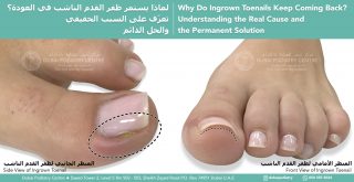
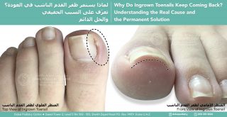
الأظافر الناشبة غالبًا ما تكون مشكلة عرض وليست مشكلة قصّ. عندما يكون ظفر القدم أعرض من الإصبع، تضغط الجوانب إلى أسفل داخل الجلد. هذا يبطئ نمو الظفر، ويؤدي إلى زيادة سماكة الجذر للظفر، ومع مرور الوقت يصبح الظفر أكثر تقوّسًا وأكثر ألمًا.

الاستئصال الجزئي للظفر هو إجراء طبي دائم في طبّ القدم، يتم فيه إزالة بضعة مليمترات فقط من حافة الظفر بعناية — بالقدر الكافي ليصبح الظفر مناسبًا للإصبع بشكل صحيح. من خلال تخفيف الضغط الجانبي، يمكن للظفر أن ينمو بحرية من الجذر مرة أخرى، مما يقلل الألم ويمنع تكرار المشكلة، مع الحفاظ على مظهر جمالي وطبيعي للظفر.

👣 عودة الوظيفة الطبيعية.
تخفيف الألم.
حل طويل الأمد.

#الظفر_الناشب #طب_القدم #صحة_القدم #العناية_بالأظافر #صحة_الأظافر #الاستئصال_الجزئي_للظفر #العناية_الطبية_بالقدم #أقدام_دون_ألم #أظافر_صحية #طب_القدم_دبي

لمزيد من المعلومات أو لحجز موعد، يرجى الاتصال بعيادتنا على
+971 4 3435390
أو عبر واتساب
+971 50 3553024


A client wants to buy some sneakers.

She went into Chanel so we advised her to take the insoles out of the shoes.

She thought she was a size 39, but size 39 was too small and so was size 40.


For more information or to book an appointment please call our clinic +971 4 3435390 or WhatsApp +971 50 3553024


.أحد العملاء تريد شراء بعض الأحذية الرياضية

.ذهبت إلى شانيل لذلك نصحناها بإخراج النعال من الحذاء

.لقد ظنت أن مقاسها ٣٩، لكن مقاس ٣٩ كان صغيرًا جدًا، وكذلك مقاس ٤٠


‎لمزيد من المعلومات أو لحجز موعد يرجى الإتصال بعيادتنا على الرقم : ٠٠٩٧١٤٣٤٣٥٣٩٠ أو التواصل على الواتساب على الرقم  ٠٠٩٧١٥٠٣٥٥٣٠٢٤


Chief Podiatrist Michelle Champlin makes a pair of corrective orthotics in carrera yellow.

تقوم رئيسة أطباء الأقدام ميشيل شامبلين بتصنيع زوج من تقويم العظام التصحيحي باللون الأصفر كاريرا


For more information or to book an appointment please call our clinic +971 4 3435390 or WhatsApp +971 50 3553024

‎لمزيد من المعلومات أو لحجز موعد يرجى الإتصال بعيادتنا على الرقم : ٠٠٩٧١٤٣٤٣٥٣٩٠ أو التواصل على الواتساب على الرقم  ٠٠٩٧١٥٠٣٥٥٣٠٢٤


Copyright Dubai Podiatry 2026. All rights reserved.

WhatsApp us