fbpx

Please wait...


BLOG

No more posts

Products Insoles Square
Dubai Podiatry Centre is the Middle East’s foremost leg and foot health experts. Specializing in biomechanics, utilizing the latest and most innovative custom orthotics amongst their array of advanced foot and leg therapies.
Prescription foot orthotics are discrete, in-shoe ‘braces’ which are designed to correct abnormal foot and leg function (including the foot, ankle, leg, knee, thigh and hip). In correcting abnormal foot and leg function, the prescription foot orthotics reduce the strain on injured structures in the foot, ankle, leg knee or hip, allowing them to heal and become non-painful. Gone are the days of ‘special shoes’ or chunky insoles.
In addition, prescription foot orthotics help prevent future problems from occurring by reducing abnormal forces on the foot and lower extremity. Custom made prescription orthotics are similar to orthodontic braces for the teeth – changing alignment, not just for cosmetic reasons but importantly to improve function, alignment, efficiency and alleviate pain. Comfort insoles bought over-the-counter from a shop or pharmacy do not correct any foot or leg malalignment but may provide some short term comfort and cushioning. Foot or leg pain should be assessed and diagnosed by a qualified Podiatrist, who complete a 4 year degree of specific medical training in the feet and legs at University (as a minimum) plus at least 2 years’ post-graduate clinical training.

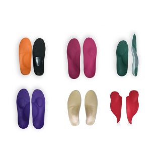
Podiatrists prescribe two main types of prescription foot orthotics for their patients: accommodative orthotics and functional/corrective foot orthotics. Both types of prescription foot orthotics are used to correct the foot posture and gait of the patient so that the pain in their foot or lower extremity will improve so that normal activities can be resumed without pain.
Custom orthotics are prescribed as a pair to allow more normal function of both feet (similar to having both the left and right wheels of a car realigned in a front end alignment).
Accommodative Foot Orthotics
Accommodative foot orthotics are used to cushion, pad or relieve pressure from a painful or injured area on the bottom of the foot. They may also be designed to try to control abnormal function of the foot. Accommodative orthotics are fabricated from a three dimensional model of the foot made by taking a plaster mold of the foot, following a thorough lower limb biomechanical assessment.
Accommodative orthotics are useful in the treatment of diabetic foot ulcerations and extensive arthritis, leg length difference and other conditions requiring offloading from certain areas of the foot. Accommodative orthotics are relatively cushioned compared to corrective orthotics. Dubai Podiatry Centre has worked to develop much more advanced accommodative orthotics than have previously been standard in healthcare, in that they are durable and much more slim fitting for many types of shoes.
Corrective Foot Orthotics
Functional, or corrective custom foot orthotics are used to correct abnormal foot and leg function and alignment. Custom corrective orthotics are used to address a wide range of foot, ankle and leg issues, including:
• Over-pronation of the sub-talar joints
• Supination
• Hallux valgus (both conservatively and post bunion surgery)
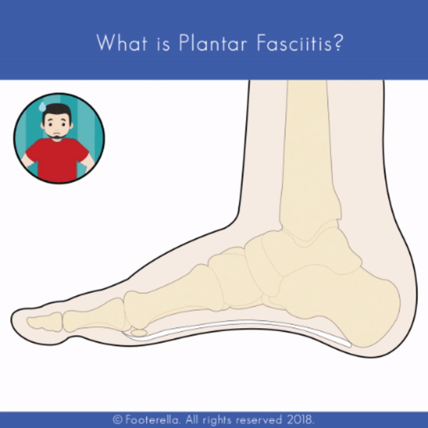
• Plantar fasciitis
• Runner’s knee
• ITB syndrome
• Ingrown toenails caused by internally rotated hallux
• Shin splints
Each pair of custom orthotics is unique and made to the patient’s individual prescription – whether to correct an ankle rolling in or out, stabilization, off-loading, raising arches, tightening the forefoot or taking strain off of the heel or plantar fascia.
The custom orthotics developed by Chief Podiatrist Michelle Champlin at Dubai Podiatry Centre are extremely slimline and easily fit into most types of shoes, including the designer dress shoes favoured at DIFC, in airline cabin shoes, ski boots, football cleats or school shoes. The advanced custom sports orthotics used by professional and amateur athletes, from cricketers to cyclists and ultra marathoners. They are fabricated from a three-dimensional model of the foot in the clinic. Almost all clinics take a mold of the patient’s foot and then outsource this off to a factory, usually in another country such as the USA, whereby these are manufactured and sent back. Dubai Podiatry Centre is unique in the Middle East in providing an end-to-end service to its patients from initial consultation through to the Podiatrist hand making the patient’s orthotics in their in-house laboratory, then fitting the orthotics to the patient. The in-house lab allows for minute adjustments within seconds to achieve very precise adjustments to within 1 degree, whilst the patient is present, saving the patient precious time.
As foot posture improves with orthotic usage, much like braces for the teeth, these can be adjusted to keep improving foot posture until foot posture goals have been achieved. Without an in-house lab, these instant changes, at the Podiatrist’s own hands would not be possible. The clinic offers a unique, bespoke and highly specialized biomechanics service to discerning patients who expect, and receive, the best results possible within the shortest timescales.

يعد مركز دبي للعناية بالاقدام من أهم الخبراء في مجال صحة الساق والقدم في الشرق الأوسط. متخصصون في الميكانيكا الحيوية، باستخدام أحدث أجهزة تقويم العظام المخصصة وأكثرها ابتكارًا من بين مجموعة علاجات القدم والساق المتقدمة.

إن أجهزة تقويم القدم الموصوفة طبيًا عبارة عن “دعامات” منفصلة توضع داخل الحذاء، وهي مصممة لتصحيح وظيفة القدم والساق غير الطبيعية (بما في ذلك القدم والكاحل والساق والركبة والفخذ والورك). من أجل تصحيح وظيفة القدم والساق غير الطبيعية، تعمل أجهزة تقويم القدم الموصوفة طبيًا على تقليل الضغط على الهياكل المصابة في القدم أو الكاحل أو ركبة الساق أو الورك، مما يسمح لها بالشفاء وتصبح غير مؤلمة. لقد ولت أيام “الأحذية الخاصة” أو النعال الداخلية المكتنزة.

بالإضافة إلى ذلك، تساعد أجهزة تقويم القدم الموصوفة طبيًا على منع حدوث مشاكل مستقبلية عن طريق تقليل القوى غير الطبيعية الموجودة على القدم والأطراف السفلية. تشبه أجهزة تقويم الأسنان الموصوفة حسب الطلب دعامات تقويم الأسنان – مما يؤدي إلى تغيير المحاذاة، ليس فقط لأسباب تجميلية ولكن من المهم تحسين الوظيفة والمحاذاة والكفاءة وتخفيف الألم. النعال الداخلية المريحة التي يتم شراؤها بدون وصفة طبية من متجر أو صيدلية لا تصحح أي سوء في القدم أو الساق ولكنها قد توفر بعض الراحة والتوسيد على المدى القصير. يجب تقييم وتشخيص آلام القدم أو الساق من قبل طبيب أقدام مؤهل، والذي أكمل درجة 4 من التدريب الطبي المحدد في القدمين والساقين في الجامعة (كحد أدنى) بالإضافة إلى تدريب سريري بعد التخرج لمدة عامين على الأقل.

يصف أطباء الأقدام نوعين رئيسيين من أجهزة تقويم القدم الموصوفة طبيًا لمرضاهم: أجهزة تقويم العظام المتكيفة وأجهزة تقويم القدم الوظيفية/التصحيحية. يتم استخدام كلا النوعين من أجهزة تقويم القدم الموصوفة طبيًا لتصحيح وضعية القدم ومشية المريض بحيث يتحسن الألم في قدمه أو الطرف السفلي بحيث يمكن استئناف الأنشطة الطبيعية دون ألم.

يتم وصف أجهزة تقويم العظام المخصصة كزوج للسماح بوظيفة طبيعية أكثر لكلا القدمين (على غرار إعادة ضبط العجلات اليسرى واليمنى للسيارة في محاذاة الواجهة الأمامية).

تقويم العظام للقدم المتكيفة

تُستخدم أجهزة تقويم القدم المتكيفة لتوسيد أو تخفيف الضغط من منطقة مؤلمة أو مصابة في الجزء السفلي من القدم. وقد تكون مصممة أيضًا لمحاولة التحكم في الوظيفة غير الطبيعية للقدم. يتم تصنيع أجهزة تقويم العظام المتكيفة من نموذج ثلاثي الأبعاد للقدم مصنوع عن طريق أخذ قالب من الجبس للقدم، بعد تقييم ميكانيكي حيوي شامل للطرف السفلي.

تعتبر أجهزة تقويم العظام التكيفية مفيدة في علاج تقرحات القدم السكرية والتهاب المفاصل الواسع النطاق واختلاف طول الساق وغيرها من الحالات التي تتطلب التفريغ من مناطق معينة من القدم. تعتبر أجهزة تقويم العظام المتكيفة مبطنة نسبيًا مقارنة بأجهزة تقويم العظام التصحيحية. لقد عمل مركز دبي لطب الأرجل على تطوير أجهزة تقويمية أكثر تقدماً بكثير مما كانت عليه في السابق في مجال الرعاية الصحية،

من حيث أنها متينة وأكثر نحافة ومناسبة للعديد من أنواع الأحذية.

تقويم العظام التصحيحية

تُستخدم أجهزة تقويم القدم الوظيفية أو التصحيحية المخصصة لتصحيح وظيفة القدم والساق غير الطبيعية ومحاذاةهما. تُستخدم أجهزة تقويم العظام التصحيحية المخصصة لمعالجة مجموعة واسعة من مشكلات القدم والكاحل والساق، بما في ذلك:

الإفراط في الكب للمفاصل تحت الكاحل

الاستلقاء

إبهام القدم الأروح (سواء بشكل محافظ أو بعد جراحة الورم)

التهاب اللفافة الأخمصية

ركبة العداء

متلازمة ITB

ظهور أظافر تحت الجلد بسبب دوران إبهام القدم إلى الداخل

جبائر قصبة الساق

كل زوج من أجهزة تقويم العظام المخصصة فريد من نوعه ويتم تصنيعه وفقًا للوصفة الطبية الفردية للمريض – سواء لتصحيح تدحرج الكاحل للداخل أو للخارج، أو التثبيت، أو التفريغ، أو رفع الأقواس، أو شد مقدمة القدم، أو إزالة الضغط من الكعب أو اللفافة الأخمصية.

إن أجهزة تقويم العظام المخصصة التي طورتها ميشيل شامبلن، كبيرة أطباء الأقدام في مركز دبي لعلاج الأرجل، رفيعة للغاية ويمكن وضعها بسهولة في معظم أنواع الأحذية، بما في ذلك الأحذية الرسمية المفضلة في مركز دبي المالي العالمي، أو في أحذية مقصورة شركات الطيران، أو أحذية التزلج، أو أحذية كرة القدم، أو الأحذية المدرسية. أجهزة تقويم العظام الرياضية المخصصة المتقدمة التي يستخدمها الرياضيون المحترفون والهواة، من لاعبي الكريكيت إلى راكبي الدراجات وسباقات الماراثون الفائقة. يتم تصنيعها من نموذج ثلاثي الأبعاد للقدم في العيادة. تأخذ جميع العيادات تقريبًا قالبًا لقدم المريض ثم تقوم بالاستعانة بمصادر خارجية لمصنع، عادة في بلد آخر مثل الولايات المتحدة الأمريكية، حيث يتم تصنيع هذه القوالب وإرسالها مرة أخرى. يعد مركز دبي لطب الأقدام فريدًا من نوعه في الشرق الأوسط من حيث تقديم خدمة شاملة لمرضاه بدءًا من الاستشارة الأولية وحتى قيام طبيب الأقدام بتصنيع أجهزة تقويم العظام للمريض في مختبره الداخلي، ثم تركيب أجهزة تقويم العظام للمريض. يسمح المختبر الداخلي بإجراء تعديلات دقيقة في غضون ثوانٍ لتحقيق تعديلات دقيقة جدًا في حدود درجة واحدة، أثناء وجود المريض، مما يوفر للمريض وقتًا ثمينًا.

مع تحسن وضعية القدم باستخدام تقويم العظام، مثل تقويم الأسنان، يمكن تعديلها لمواصلة تحسين وضعية القدم حتى يتم تحقيق أهداف وضعية القدم. وبدون وجود مختبر داخلي، فإن هذه التغييرات الفورية، على يد طبيب الأقدام، لن تكون ممكنة. تقدم العيادة خدمة ميكانيكا حيوية فريدة ومخصصة وعالية التخصص للمرضى المميزين الذين يتوقعون ويحصلون على أفضل النتائج الممكنة في أقصر الفترات الزمنية.


Written by Michelle Champlin BSc Pod., M.Ch.S., S.R., Ch., (UK)
Most of us experience swollen feet at some point. If it lasts more than a couple of days, or is accompanied by other signs such as pain, persistent cough, change in skin colour or sensation, or is in only one leg, you should see your Doctor or GP straight away.
1. Cold Feet
While it is possible to simply have cold sensations without any serious underlying cause, chronically cold toes can be a sign of impaired blood circulation. Smoking and smoking-related conditions like COPD can reduce the lungs’ ability to fully absorb oxygen, causing reduced oxygen levels in the blood. Additionally, peripheral vascular disease (PVD) and some other forms of heart disease can cause your arteries to narrow, which can impede the flow of blood throughout the body. Extremities like hands and feet are particularly susceptible to poor circulation, which may manifest itself through cold sensations. (It’s one of the reasons why a Podiatrist tests circulation in the feet of a Diabetic regularly – to pick up any changes early before it affects the rest of the body / organs). If you notice that your feet are almost always cold or numb, consult your Doctor or Podiatrist to figure out whether there are any underlying medical problems.
2. Itchy Feet
The most common cause of itchy, scaly skin on the feet is a fungal infection such as athlete’s foot, which is extremely common in hot, sandy climates such as the UAE. These infections thrive in damp, dark environments, usually beginning between the toes and the risk is raised by enclosed shoes of synthetic, non breathable material and sweaty feet. While athlete’s foot is contagious, it is relatively easy for a Podiatrist to treat. Michelle Champlin, Chief Podiatrist at Dubai Podiatry Centre advises a visit to your Podiatrist to remove any hard skin (the fungal spores feed on keratin in dead skin), check your toenails in case it has spread, and to provide a strong once off topical anti-fungal along with follow-up at home medication such as antifungal powder and spray to treat your socks and shoes with to prevent reinfection. More of Mrs Champlin’s tips on getting rid of fungal feet can be found here. In severe cases, oral antifungals may be prescribed. In some cases, itchy feet may also result from an allergic reaction to skin creams or from an immune condition like psoriasis or eczema.
3. Skin Discolouration
A rash or fungal infection like athlete’s foot commonly leads to scaly, reddish skin, or also white, powdery looking skin. Many other conditions also result in discoloration of the feet. One condition, known as Raynaud’s phenomenon, is characterized by a sequence of color changes in the skin as a response to cold or stress. During a Raynaud’s flare up, the affected areas (usually feet or hands) turn white initially as the arteries begin to narrow and blood flow is reduced. They may start to feel cold or numb and eventually turn blue as blood flow is further restricted. Sometimes, as circulation improves, the skin may turn red before finally returning to normal. Raynaud’s occurs most frequently without an underlying disease. In some cases, however, Raynaud’s may be an early sign of a more serious condition like rheumatoid arthritis or lupus.
4. Swollen Feet
Most people experience swelling in their feet (called edema) at one point in their lives and in hot climates such as the UAE mild swelling of the feet is a normal consequence to a hot climate. Often, the cause of the swelling is due to something as simple and temporary as standing for a long period of time, wearing tight footwear, or flying. In these cases, simple measures such as following in-flight exercises, walking around the cabin where possible, wearing flight socks and comfortable footwear can all help. However, if the swelling lasts for more than a couple of days, it could be a sign of an injury such as an ankle sprain or break, or more serious medical condition. Fluids may build up in your legs and cause swelling as a result of congestive heart failure, kidney disease or even as a side effect of prescription medications for diabetes and high blood pressure. Swelling may also result from inflammation stemming from rheumatoid arthritis or osteoarthritis. It is important to seek emergency medical attention if swelling is accompanied by symptoms such as chest pain, difficulty breathing or dizziness, as these could indicate a blood clot or another serious heart condition. It is important whether the swelling is to one or both feet or legs – swelling to one leg only can be a sign of DVT/blood clot and emergency medical care should be sought. Other causes of swollen feet include pregnancy, lymphedema or gout. In any case, see your GP or Podiatrist for unexplained swelling in your feet that does not go away.
5. Burning Feet
The sensation of burning feet can vary from mild (numbness and tingling) to severe (distracting pain that disrupts your sleep). Something as simple as tired feet or a common infection like athlete’s foot can cause short-term symptoms like burning or tingling. In more severe cases, burning in the feet could be a sign of nerve damage from diabetes or a circulatory condition known as peripheral vascular disease (PVD). According to the International Diabetes Federation, as of 2014 10.7% of the population in the UAE had been diagnosed with diabetes – that’s 803,900 people, with this number on the increase. Seek medical attention from your GP or Podiatrist if the burning sensation lasts for several weeks, if symptoms become more intense and painful or if you start losing feeling in your toes or feet. In the meantime, rest and elevate your feet to ease symptoms. Additionally, bathing your feet in cold water can also reduce pain and burning. Burning feet may signal that your diabetes management / medication needs to be altered and early intervention from your Doctor or endocrinologist is crucial. Burning-like sensation can also be caused by nerve entrapment (neuromas).
If you are worried about any change in your feet, contact the expert Podiatrists at Dubai Podiatry Centre without delay. Contact +971 4 3435390.


٥ علامات على الأقدام لا يجب أن تتجاهلها أبدًا

بقلم ميشيل شامبلين

يعاني معظمنا من تورم القدمين في مرحلة ما. إذا استمرت أكثر من يومين، أو كانت مصحوبة بعلامات أخرى مثل الألم، أو السعال المستمر، أو تغير في لون الجلد أو الإحساس، أو كانت في ساق واحدة فقط، فيجب عليك مراجعة طبيبك أو طبيبك العام على الفور.

قدم باردة

في حين أنه من الممكن أن تشعر ببساطة بالأحاسيس الباردة دون أي سبب أساسي خطير، إلا أن برودة أصابع القدم المزمنة يمكن أن تكون علامة على ضعف الدورة الدموية. التدخين والحالات المرتبطة بالتدخين مثل مرض الانسداد الرئوي المزمن يمكن أن تقلل من قدرة الرئتين على امتصاص الأكسجين بشكل كامل، مما يسبب انخفاض مستويات الأكسجين في الدم. بالإضافة إلى ذلك، يمكن أن يتسبب مرض الأوعية الدموية الطرفية (PVD) وبعض الأشكال الأخرى من أمراض القلب في تضييق الشرايين، مما قد يعيق تدفق الدم في جميع أنحاء الجسم. الأطراف مثل اليدين والقدمين معرضة بشكل خاص لضعف الدورة الدموية، والذي قد يتجلى من خلال الأحاسيس الباردة. (إنه أحد الأسباب التي تجعل طبيب الأقدام يقوم باختبار الدورة الدموية في قدمي مريض السكري بانتظام – لاكتشاف أي تغييرات مبكرًا قبل أن تؤثر على بقية الجسم / الأعضاء). إذا لاحظت أن قدميك دائمًا ما تكون باردة أو مخدرة، استشر طبيبك أو طبيب الأقدام لمعرفة ما إذا كانت هناك أي مشاكل طبية كامنة.

حكة في القدمين

السبب الأكثر شيوعًا لحكة الجلد المتقشر في القدمين هو العدوى الفطرية مثل قدم الرياضي، وهو أمر شائع للغاية في المناخات الرملية الحارة مثل دولة الإمارات العربية المتحدة. تزدهر هذه العدوى في البيئات الرطبة المظلمة، وعادةً ما تبدأ بين أصابع القدم، ويزداد الخطر عند ارتداء الأحذية المغلقة المصنوعة من مواد اصطناعية غير قابلة للتنفس والأقدام المتعرقة. على الرغم من أن قدم الرياضي معدية، إلا أنه من السهل نسبيًا علاجها بواسطة طبيب الأقدام. تنصح ميشيل شامبلين، رئيسة أطباء الأقدام في مركز دبي لعلاج الأقدام بزيارة طبيب الأقدام الخاص بك لإزالة أي جلد صلب (تتغذى الجراثيم الفطرية على الكيراتين الموجود في الجلد الميت)، وفحص أظافر قدميك في حالة انتشارها، وتوفير مضاد موضعي قوي لمرة واحدة. – الفطريات مع متابعة الأدوية المنزلية مثل المسحوق المضاد للفطريات والرذاذ لعلاج جواربك وأحذيتك لمنع الإصابة مرة أخرى. يمكن العثور على المزيد من نصائح السيدة شامبلن حول التخلص من فطريات القدم هنا. في الحالات الشديدة، يمكن وصف مضادات الفطريات عن طريق الفم. في بعض الحالات، قد تنجم حكة القدم أيضًا عن رد فعل تحسسي تجاه كريمات الجلد أو عن حالة مناعية مثل الصدفية أو الأكزيما.

تغير لون الجلد. ٣

عادةً ما يؤدي الطفح الجلدي أو العدوى الفطرية، مثل قدم الرياضي، إلى ظهور جلد متقشر ومحمر، أو أيضًا بشرة بيضاء ذات مظهر مسحوقي. العديد من الحالات الأخرى تؤدي أيضًا إلى تغير لون القدمين. إحدى الحالات، المعروفة باسم ظاهرة رينود، تتميز بسلسلة من التغيرات اللونية في الجلد كرد فعل للبرد أو الإجهاد. أثناء نوبة رينود، تتحول المناطق المصابة (عادةً القدمين أو اليدين) إلى اللون الأبيض في البداية عندما تبدأ الشرايين في التضييق ويقل تدفق الدم. قد يبدأون في الشعور بالبرد أو الخدر ويتحولون في النهاية إلى اللون الأزرق بسبب تقييد تدفق الدم بشكل أكبر. في بعض الأحيان، مع تحسن الدورة الدموية، قد يتحول الجلد إلى اللون الأحمر قبل أن يعود أخيرًا إلى طبيعته. يحدث مرض رينود في أغلب الأحيان دون وجود مرض أساسي. ومع ذلك، في بعض الحالات، قد يكون مرض رينود علامة مبكرة على حالة أكثر خطورة مثل التهاب المفاصل الروماتويدي أو الذئبة.

تورم القدمين

يعاني معظم الأشخاص من تورم في أقدامهم (يسمى الوذمة) في مرحلة ما من حياتهم، وفي المناخات الحارة مثل دولة الإمارات العربية المتحدة، يعد التورم الخفيف في القدمين نتيجة طبيعية للمناخ الحار. في كثير من الأحيان، يرجع سبب التورم إلى شيء بسيط ومؤقت مثل الوقوف لفترة طويلة من الزمن، أو ارتداء الأحذية الضيقة، أو الطيران. في هذه الحالات، يمكن أن تساعد الإجراءات البسيطة مثل اتباع التمارين أثناء الرحلة، والتجول في المقصورة حيثما أمكن ذلك، وارتداء جوارب الطيران والأحذية المريحة. ومع ذلك، إذا استمر التورم لأكثر من يومين، فقد يكون ذلك علامة على وجود إصابة مثل التواء أو كسر في الكاحل، أو حالة طبية أكثر خطورة. قد تتراكم السوائل في ساقيك وتسبب التورم نتيجة لقصور القلب الاحتقاني أو أمراض الكلى أو حتى كأثر جانبي للأدوية الموصوفة لمرض السكري وارتفاع ضغط الدم. قد ينجم التورم أيضًا عن التهاب ناجم عن التهاب المفاصل الروماتويدي أو التهاب المفاصل العظمي. من المهم طلب العناية الطبية الطارئة إذا كان التورم مصحوبًا بأعراض مثل ألم في الصدر أو صعوبة في التنفس أو دوخة، لأن هذه قد تشير إلى جلطة دموية أو حالة قلبية خطيرة أخرى. من المهم ما إذا كان التورم في إحدى القدمين أو الساقين أو كلتيهما – يمكن أن يكون التورم في ساق واحدة فقط علامة على الإصابة بجلطات الأوردة العميقة / جلطة دموية ويجب طلب الرعاية الطبية الطارئة. تشمل الأسباب الأخرى لتورم القدمين الحمل أو الوذمة اللمفية أو النقرس. على أية حال، راجع طبيبك العام أو طبيب الأقدام لمعرفة التورم غير المبرر في قدميك والذي لا يختفي.

حرق القدمين . ٥

يمكن أن يختلف الإحساس بحرقان القدمين من خفيف (خدر ووخز) إلى شديد (ألم مشتت يعطل نومك). يمكن لشيء بسيط مثل الأقدام المتعبة أو عدوى شائعة مثل قدم الرياضي أن يسبب أعراضًا قصيرة المدى مثل الحرق أو الوخز. في الحالات الأكثر شدة، يمكن أن يكون حرق القدمين علامة على تلف الأعصاب بسبب مرض السكري أو حالة الدورة الدموية المعروفة باسم مرض الأوعية الدموية الطرفية (PVD). وفقًا للاتحاد الدولي للسكري، اعتبارًا من عام ٢٠١٤، تم تشخيص إصابة ١٠.٧% من السكان في دولة الإمارات العربية المتحدة بمرض السكري – أي ٨٠٣،٩٠٠ شخص، وهذا العدد في ازدياد. اطلب العناية الطبية من طبيبك العام أو طبيب الأقدام إذا استمر الإحساس بالحرقان لعدة أسابيع، أو إذا أصبحت الأعراض أكثر شدة وألمًا، أو إذا بدأت تفقد الإحساس في أصابع قدميك أو قدميك. في هذه الأثناء، استرح وارفع قدميك لتخفيف الأعراض. بالإضافة إلى ذلك، فإن تحميم قدميك في الماء البارد يمكن أن يقلل أيضًا من الألم والحرقان. قد يشير حرق القدمين إلى أن إدارة مرض السكري / الدواء الخاص بك يحتاج إلى تغيير وأن التدخل المبكر من طبيبك أو أخصائي الغدد الصماء أمر بالغ الأهمية. يمكن أيضًا أن يحدث الإحساس الشبيه بالحرقان بسبب انحباس العصب (الأورام العصبية).

إذا كنت قلقًا بشأن أي تغيير في قدميك، تواصل مع خبراء طب الأقدام في مركز دبي للعناية بالأقدام دون تأخير. اتصل بالرقم ٠٤٣٤٣٥٣٩٠


https://dubaipodiatry.com/wp-content/uploads/2017/06/ezgif.com-video-to-gif-24.gif

Plantar heel pain is one of the most common musculoskeletal conditions that is seen at the specialist foot clinic on Sheikh Zayed Road, Dubai Podiatry Centre. It can cause significant pain, especially in the mornings and can cause significant impact on walking (causing a limp), productivity and general quality of life.

Chief Podiatrist Michelle Champlin explains, “Alleviating plantar fasciitis is one of the most rewarding treatments that we provide – from identifying any underlying biomechanical cause, to making just the right bespoke custom orthotics for the patient to alleviate it. It is fabulous to alleviate pain that patients may have been suffering with for weeks, months or even years and who may have tried all sorts of therapies to no avail.”

يعد ألم الكعب الأخمصي أحد أكثر حالات الجهاز العضلي الهيكلي شيوعًا والتي يتم ملاحظتها في عيادة القدم المتخصصة في شارع الشيخ زايد، مركز دبي لعلاج الأرجل. يمكن أن يسبب ألمًا كبيرًا، خاصة في الصباح ويمكن أن يسبب تأثيرًا كبيرًا على المشي (مما يسبب العرج)، والإنتاجية ونوعية الحياة بشكل عام.

تشرح رئيسة أطباء الأقدام ميشيل شامبلين قائلة: “يعد تخفيف التهاب اللفافة الأخمصية واحدًا من أكثر العلاجات المجزية التي نقدمها – بدءًا من تحديد أي سبب ميكانيكي حيوي أساسي، إلى صنع أجهزة تقويم العظام المخصصة والمناسبة للمريض للتخفيف من حدتها. إنه أمر رائع لتخفيف الألم الذي قد يعاني منه المرضى لأسابيع أو أشهر أو حتى سنوات والذين ربما جربوا جميع أنواع العلاجات دون جدوى.


Patients have frequently tried interventions such as plantar arch strapping, over-the-counter arch supports and heel cups before they come to see Dubai Podiatry Centre. The chronic pain can be debilitating and affect everything from your ability to walk around the mall or stand at work.

لقد جرب المرضى في كثير من الأحيان تدخلات مثل ربط القوس الأخمصي، ودعامات القوس المتاحة دون وصفة طبية وأكواب الكعب قبل أن يأتوا لزيارة مركز دبي لعلاج الأرجل. يمكن أن يكون الألم المزمن منهكًا ويؤثر على كل شيء بدءًا من قدرتك على التجول في المركز التجاري أو الوقوف في العمل


How Do The Biomechanics Of Plantar Heel Pain Relate To Foot Orthotic Therapy?

Heel pain can have a number of causes including plantar fasciitis, subcalcaneal bursitis, neuritis, medial arch pain, subcalcaneal pain, stone bruise, calcaneal periostitis, subcalcaneal spur or heel bone fracture. However, the majority of patients exhibiting plantar heel pain have pathology related to injury of the plantar fascia i.e. plantar fasciitis is the most common cause of heel pain.

There are many risk factors for plantar heel pain, including increased body mass index (BMI), limited ankle joint dorsiflexion (e.g. ankle equinus), heel spur, leg length discrepancy, diminished heel fatty pad thickness, pes planus (flat feet), pes cavus (high arches), excess overpronation and hallux limitus or hallux rigidus (reduced range of movement in the big toe joint).

The plantar fascia connects your heel to your forefoot, providing support like the bottom beam of a roof truss

كيف ترتبط الميكانيكا الحيوية لألم الكعب الأخمصي بعلاج تقويم العظام للقدم؟

يمكن أن يكون لألم الكعب عدد من الأسباب بما في ذلك التهاب اللفافة الأخمصية، والتهاب الجراب تحت العقبي، والتهاب العصب، وألم القوس الإنسي، وألم تحت العقبي، وكدمة الحجر، والتهاب السمحاق العقبي، والمهماز تحت العقبي أو كسر عظم الكعب. ومع ذلك، فإن غالبية المرضى الذين يعانون من آلام الكعب الأخمصي لديهم أمراض مرتبطة بإصابة اللفافة الأخمصية، أي أن التهاب اللفافة الأخمصية هو السبب الأكثر شيوعًا لألم الكعب.

هناك العديد من عوامل الخطر لألم الكعب الأخمصي، بما في ذلك زيادة مؤشر كتلة الجسم (BMI)، محدودية عطف ظهري مفصل الكاحل (مثل الاعتدال في الكاحل)، مهماز الكعب، التناقض في طول الساق، انخفاض سمك الطبقة الدهنية للكعب، القدم المسطحة، pes. التجويف (الأقواس العالية)، الكب الزائد الزائد وإبهام القدم المحدود أو إبهام القدم الجامد (نطاق منخفض من الحركة في مفصل إصبع القدم الكبير).

تربط اللفافة الأخمصية كعبك بمقدمة قدمك، مما يوفر الدعم مثل العارضة السفلية لدعامات السقف


https://dubaipodiatry.com/wp-content/uploads/2017/06/ezgif.com-video-to-gif-22-1.gif

What Are The Potential Biomechanical Effects Of Foot Orthotics On The Plantar Fascia?

The plantar fascia is a very strong but non-stretchy connective tissue between your heel and forefoot, also known as the plantar aponeurosis. It acts almost like an ‘emergency band’ which carries the bulk of load during standing and walking, maintaining the structural integrity of the arch of the foot when the foot’s other tendons, ligaments and muscles do not support foot posture as they should.

At Dubai Podiatry Centre, the Podiatrists think more from a structural engineering standpoint, comparing foot structure to both a structured beam as well as a truss, stabilized by a “tie,” which is the plantar fascia. When weight-bearing, the plantar fascia prevents the ends of the arch from elongating. This mechanism helps explain the ‘biomechanics’ of using arch supporting custom orthotics to treat plantar fasciitis.

A study by Kogler et al found that orthotics significantly reduced strain in the plantar fascia. However, it is important to note that only those orthoses that had significant contour under the “apical” portion of the arch (i.e. talonavicular joint) provide benefit, which is one of the reasons why Dubai Podiatry Centre provides custom corrective orthotics made by the Podiatrists in their in-house lab, from a mould of the patient’s feet. This ensures an exact shape is tailor made with the precise prescription applied to correct foot posture as required – no two feet are the same and different prescriptions, just like braces for the teeth, are required for each patient.

Foot orthotics provide immediate, intermediate and long-term relief of plantar heel pain with significant pain relief normally felt within the first six weeks.

ما هي التأثيرات الميكانيكية الحيوية المحتملة لتقويم القدم على اللفافة الأخمصية؟

اللفافة الأخمصية عبارة عن نسيج ضام قوي جدًا ولكنه غير قابل للتمدد بين الكعب ومقدمة القدم، ويُعرف أيضًا باسم السفاق الأخمصي. إنه يعمل تقريبًا مثل “حزام الطوارئ” الذي يحمل الجزء الأكبر من الحمل أثناء الوقوف والمشي، ويحافظ على السلامة الهيكلية لقوس القدم عندما لا تدعم الأوتار والأربطة والعضلات الأخرى في القدم وضعية القدم كما ينبغي.

في مركز دبي لعلاج الأقدام، يفكر أطباء الأقدام أكثر من وجهة نظر الهندسة الإنشائية، حيث يقارنون هيكل القدم بكل من العارضة الهيكلية وكذلك الجمالون، المثبت بواسطة “ربطة عنق”، وهي اللفافة الأخمصية. عند حمل الوزن، تمنع اللفافة الأخمصية أطراف القوس من الاستطالة. تساعد هذه الآلية في شرح “الميكانيكا الحيوية” لاستخدام تقويم العظام المخصص الداعم للقوس لعلاج التهاب اللفافة الأخمصية.

وجدت دراسة أجراها كوجلر وآخرون أن تقويم العظام يقلل بشكل كبير من إجهاد اللفافة الأخمصية. ومع ذلك، من المهم ملاحظة أن أجهزة تقويم العظام التي لها محيط كبير تحت الجزء “القمي” من القوس (أي المفصل الكاحلي الزورقي) فقط هي التي توفر الفائدة، وهو أحد الأسباب التي تجعل مركز دبي لطب الأرجل يقدم أجهزة تقويمية تصحيحية مخصصة من صنع أطباء الأقدام. في معملهم الداخلي، من قالب قدم المريض. وهذا يضمن أن يتم تصميم الشكل الدقيق وفقًا للوصفة الطبية الدقيقة المطبقة لتصحيح وضع القدم كما هو مطلوب – لا يوجد قدمان متماثلتان ويتطلب الأمر وصفات مختلفة، تمامًا مثل تقويم الأسنان، لكل مريض.

توفر أجهزة تقويم القدم راحة فورية ومتوسطة وطويلة الأمد من آلام الكعب الأخمصي مع تخفيف كبير للألم يتم الشعور به عادةً خلال الأسابيع الستة الأولى.


BOOK AN APPOINTMENT

Send us a message on our WhatsApp for appointments and other queries.
WhatsApp : +971 50 331 4978 | Call: +971  4  343 5390


Written by Michelle Champlin BSc Pod., M.Ch.S., S.R., Ch., (UK) Over-pronation & Your Knees The subtalar joint, also known as the talocalcaneal joint, is one of the 33 joints in the foot. It is located at the meeting point of the talus bone and the calcaneus (heel bone), below the ankle joint. It is this joint that can cause the foot and ankle to ‘roll in’ giving the appearance of flat feet, called ‘over-pronation’. The subtalar joint movement and range of motion is transferred up into the leg by rotating the tibia bone and also forward into the foot, controlling the locking and unlocking of another foot joint called the midtarsal joint, all necessary for normal standing, walking and running. Over-pronation at the ankle can cause excess strain and pain to the knee Over-pronation of the Subtalar Joint & the Knee The effect of the subtalar joint on the knee as it causes the internal and external rotation of the tibia is a change in the alignment of the knee patella and the patella tendon. When the subtalar joint is misaligned, this rotation of the tibia causes the patella to ride abnormally with the articulation with the femur in the thigh. This is a common root cause of a knee condition called chrondomalacia patella, advises Chief Podiatrist Michelle Champlin. Additionally, with excessive rotation of the tibia, the collateral knee ligaments are strained. Left untreated, this can result in arthroscopic knee surgeries to correct the damage caused ultimately by a misaligned subtalar joint in the foot. Even after knee surgery, unless the subtalar joint over-pronation is addressed, the knee joint degeneration will continue. Other issues caused by over-pronation of the subtalar joint include ‘acquired’ flat feet. This is a biomechanics issue that was not present at birth and gets worse over time. However, this can generally be corrected. Travelling up the body, joints at the ankles, knees and hips can be affected. The over-pronating subtalar joint can also cause problems in the smaller joints of the forefoot and toes, leading to bunions, bunionettes, neuromas and hammer/claw toes. Diagnosis The most accurate diagnosis of subtalar joint over-pronation is made by a skilled Podiatrist observing the patient walking, standing and non weight bearing, as well as performing a hands on evaluation and manipulation of the foot and ankle. Observation of the foot in a walking examination is most reliable. All together, this is called a lower limb biomechanical assessment. Treatment Over-pronation is best treated early. There is no recommended home treatment other than the general avoidance of prolonged weight bearing in non-supportive footwear such as flip flops or ballet pumps until the patient can be assessed and treated by a Podiatrist. Treatment with custom-molded foot orthotics worn in regular footwear or sports shoes is critical to maintain stability of the foot and ankle and to improve the foot posture. The purpose of the orthotics is to get the subtalar joint functioning properly on its own again by tightening and toning the supporting ligaments, tendons and muscles. As with all progressive medical conditions, it is best to start treatment early. Chief Podiatrist Michelle Champlin of Dubai Podiatry Centre recommends that all school age children are screened at age 4-5 initially for this reason. Orthotics Custom orthotic treatment directed at alleviating symptoms such as arch, knee or hip pain and slowing or halting the progression is the goal. The custom orthotics must be made to the unique specifications of the patient if they are to achieve the desired results. They must also be constructed to fit in regular footwear, with Dubai Podiatry Centre’s orthotics being on average only several millimeters thick, with a variety of custom designs to accommodate even slim fitting dress shoes, football cleats or regular running shoes. It is important to the experts at Dubai Podiatry Centre that the orthotics are fitted properly into the patient’s footwear, which is one of the reasons they see the patients with their footwear and fit the orthotics into it, having the patient try the orthotics on and walk in them. To capture the correct prescription of the orthotics, Podiatrists must have a through understanding of lower leg function. At Dubai Podiatry Centre, the Podiatrists take a specific mold of the patient’s feet, in a specific position and based upon their measurements of the subtalar joints and midtarsal joints of the foot and their ranges of motion. The Podiatrists take these measurements and then evaluate them relative to a weight bearing gait analysis. Thorough biomechanical assessment takes place in the Dubai clinic on Sheikh Zayed Road, where the Podiatrist has access to their detailed instruments, materials and equipment. To improve results for the patient further, the Podiatrists at Dubai Podiatry Centre have access to their own in-clinic orthotic laboratory. The Podiatrists understand the proper construction of a corrective orthotic and hand make their own patients’ orthotics, ensuring complete control over the patient’s treatment and the highest quality orthotic. A critical element in the successful treatment of knee and foot issues with custom corrective orthotics is for the treating Podiatrist, who has the training, knowledge and experience in the treatment of these issues, to maintain total control of the casting, measuring and fabrication of the orthotics. Health Insurance & Orthotics
Occasionally, should a patient’s the health insurance plan set their fees below a level to reimburse the patient for their Podiatrist’s time, expertise and laboratory costs, then the patient may delay treatment or seek alternative insoles or providers not equipped to meet the patients needs, which in time usually results in the problem not being properly corrected, further degeneration of the knee or other affected joints and ultimately a more costly financial outcome. Dubai Podiatry Centre’s Podiatrists provide all patients with a detailed medical report outlining expected costs for patients to send to their Insurer to seek pre-authorisation and to understand the extent of their reimbursement and coverage. If you are concerned that your feet appear ‘flat’ or have associated knee, ankle or hip pain see a specialist biomechanics Podiatrist, who has an in-house clinic to make corrective orthotics and provide important follow up monitoring to change the prescription and monitor improvement. Consultation with the expert Podiatrists at Dubai Podiatry Centre can be made on +971 4 3435390. The navicular bone in humans is one of the tarsal bones, found in the foot. Its name comes from the bone’s resemblance to a small boat, caused by its very concave proximal articular surface. The navicular bone is located on the medial (upper inside) side of the foot, and connects the talus (ankle bone) behind it, with the three cuneiform bones in front of it, and to the side with the cuboid. The navicular bone forms a very important joint – the talocalcaneonavicular joint – with the talus (ankle bone) and calcaneum (heel bone). This joint transmits the entire weight of the body through the foot. The navicular bone, in orange, sits with the cuneiform bones in front of it and talus bone behind it It is the last of the foot’s 26 bones to start ossification (laying down bone) and does not tend to do so until the end of the second year in girls and the beginning of the fourth year in boys, although this can vary from person to person. The tibialis posterior is the only muscle that attaches to the navicular bone. Navicular Injuries Injuring the navicular bone is relatively rare. Fracture of the navicular though can occur usually in athletes whilst kicking, sprinting, twisting, or falling. This will cause pain and limping. Treatment options for injury to the navicular include utilizing a non-weight bearing aircast to hold the bone stable so it can mend. If surgery is recommended, screws will be placed into the bone to help secure it. The patient will wear a cast after the surgery. In many cases, pain associated with injuries to the bone will lessen, and later return when that part of the foot is manipulated. As a result, the Podiatrist or Surgeon will manually check the navicular following treatment to see if there is pain when the top of the foot is manipulated. The other problem that can affect the navicular bone is ‘accessory navicular syndrome’. Occasionally, some people are born with an extra bone or piece of cartilage, just above the arch on the inside of the foot. This may cause no symptoms and be entirely harmless. In some cases, the bone or the posterior tibial tendon attaching to the navicular becomes inflamed and painful. This can be caused by a trauma such as an ankle sprain, footwear rubbing on this bone or being extremely active. Many people with this extra bone will also have flat feet, putting more strain on the tendon in this area also. This syndrome tends to develop in teenage years. Treatment
Following a thorough biomechanical assessment and medical history, the Podiatrist may recommend a range of treatments such as immobilization with an aircast, icing, NSAIDs for pain and inflammation, or custom orthotics to raise the arch and correct foot posture. If conservative treatment options do not provide relief, If you or your child experience pain, swelling or irritation of your foot arch, contact the foot and ankle specialist clinic, Dubai Podiatry Centre on +971 4 3435390 for rapid diagnosis and treatment.  

بقلم ميشيل شامبلين

الإفراط في الميلان وركبتيك

المفصل تحت الكاحل، المعروف أيضًا باسم المفصل الكاحلي العقبي، هو واحد من 33 مفصلًا في القدم. وهو يقع عند نقطة التقاء عظم الكاحل والعقب (عظم الكعب)، أسفل مفصل الكاحل. هذا المفصل هو الذي يمكن أن يتسبب في “تدحرج” القدم والكاحل مما يعطي مظهر القدم المسطحة، وهو ما يسمى “الميلان الزائد“.

يتم نقل حركة المفصل تحت الكاحل ونطاق الحركة إلى الأعلى إلى الساق عن طريق تدوير عظم الظنبوب وإلى الأمام أيضًا في القدم، للتحكم في قفل وفتح مفصل قدم آخر يسمى المفصل الوسطي، وكلها ضرورية للوقوف والمشي والجري بشكل طبيعي.

الإفراط في الميلان في الكاحل يمكن أن يسبب الضغط الزائد والألم في الركبة

الإفراط في الميلان للمفصل تحت الكاحل والركبة

تأثير المفصل تحت الكاحل على الركبة حيث أنه يسبب الدوران الداخلي والخارجي للظنبوب هو تغيير في محاذاة رضفة الركبة ووتر الرضفة. عندما يكون المفصل تحت الكاحل منحرفًا، يؤدي هذا الدوران للظنبوب إلى تحرك الرضفة بشكل غير طبيعي مع المفصل مع عظم الفخذ في الفخذ.

هذا هو السبب الجذري الشائع لحالة الركبة التي تسمى تلين الرضفة، كما تنصح رئيسة أطباء الأقدام ميشيل شامبلين. بالإضافة إلى ذلك، مع الدوران المفرط لعظم الساق، تتوتر الأربطة الجانبية للركبة. إذا تُرك هذا الأمر دون علاج، فقد يؤدي ذلك إلى إجراء عمليات جراحية بالمنظار في الركبة لتصحيح الضرر الناتج في النهاية عن محاذاة المفصل تحت الكاحل بشكل غير صحيح في القدم. حتى بعد جراحة الركبة، ما لم تتم معالجة فرط كب المفصل تحت الكاحل، فإن انحطاط مفصل الركبة سيستمر

تشمل المشكلات الأخرى الناجمة عن الإفراط في كب المفصل تحت الكاحل الأقدام المسطحة “المكتسبة”. هذه مشكلة في الميكانيكا الحيوية لم تكن موجودة عند الولادة وتزداد سوءًا بمرور الوقت. ومع ذلك، يمكن تصحيح هذا بشكل عام. أثناء السفر إلى أعلى الجسم، يمكن أن تتأثر المفاصل عند الكاحلين والركبتين والوركين. يمكن أيضًا أن يسبب المفصل تحت الكاحل المفرط النطق مشاكل في المفاصل الأصغر في مقدمة القدم وأصابع القدم، مما يؤدي إلى الأورام والأورام العصبية وأصابع القدم المطرقية/المخلبية.

تشخبص

يتم إجراء التشخيص الأكثر دقة للكب الزائد للمفصل تحت الكاحل من قبل طبيب أقدام ماهر يراقب المريض أثناء المشي والوقوف وعدم تحمل الوزن، بالإضافة إلى إجراء تقييم عملي والتلاعب بالقدم والكاحل. تعتبر مراقبة القدم أثناء فحص المشي هي الأكثر موثوقية. يُطلق على هذا معًا تقييم الميكانيكا الحيوية للطرف السفلي.

علاج

من الأفضل علاج الإفراط في الكب مبكرًا. لا يوجد علاج منزلي موصى به بخلاف التجنب العام لتحمل الوزن لفترة طويلة بارتداء أحذية غير داعمة مثل الصنادل أو أحذية الباليه حتى يمكن تقييم المريض وعلاجه بواسطة طبيب الأقدام.

يعد العلاج باستخدام أجهزة تقويم القدم المصممة خصيصًا والتي يتم ارتداؤها في الأحذية العادية أو الأحذية الرياضية أمرًا بالغ الأهمية للحفاظ على ثبات القدم والكاحل وتحسين وضعية القدم. الغرض من تقويم العظام هو جعل المفصل تحت الكاحل يعمل بشكل صحيح من تلقاء نفسه مرة أخرى عن طريق شد وتنعيم الأربطة والأوتار والعضلات الداعمة.

كما هو الحال مع جميع الحالات الطبية المتقدمة، فمن الأفضل أن تبدأ العلاج في وقت مبكر. توصي رئيسة أطباء الأقدام ميشيل شامبلين من مركز دبي لعلاج الأرجل بإجراء فحص لجميع الأطفال في سن المدرسة في سن 4-5 سنوات في البداية لهذا السبب

تقويم العظام

الهدف هو العلاج التقويمي المخصص الذي يهدف إلى تخفيف الأعراض مثل آلام القوس أو الركبة أو الورك وإبطاء أو إيقاف التقدم.

يجب أن يتم تصنيع أجهزة تقويم العظام المخصصة وفقًا للمواصفات الفريدة للمريض إذا أراد تحقيق النتائج المرجوة. ويجب أيضًا تصميمها لتناسب الأحذية العادية، حيث يبلغ متوسط سمك أجهزة تقويم العظام في مركز دبي لطب الأقدام عدة ملليمترات فقط، مع مجموعة متنوعة من التصميمات المخصصة لاستيعاب حتى الأحذية الرسمية الضيقة، أو أحذية كرة القدم، أو أحذية الجري العادية. من المهم للخبراء في مركز دبي لطب الأرجل أن يتم تركيب أجهزة تقويم العظام بشكل صحيح في أحذية المريض، وهذا أحد الأسباب التي تجعلهم يرون المرضى بأحذيتهم ويضعون أجهزة تقويم العظام فيها، مما يجعل المريض يجرب جهاز تقويم العظام ويمشي. فيهم.

للحصول على الوصفة الطبية الصحيحة لتقويم العظام، يجب أن يكون لدى أطباء الأقدام فهم شامل لوظيفة الجزء السفلي من الساق. في مركز دبي لعلاج الأرجل، يأخذ أطباء الأرجل قالبًا محددًا لقدم المريض، في وضع محدد وبناءً على قياساتهم للمفاصل تحت الكاحل والمفاصل الوسطى للقدم ونطاقات حركتها. يأخذ أطباء الأقدام هذه القياسات ثم يقومون بتقييمها مقارنة بتحليل المشية الذي يحمل الوزن.

يتم إجراء تقييم ميكانيكي حيوي شامل في عيادة دبي الواقعة على طريق الشيخ زايد، حيث يمكن لطبيب الأقدام الوصول إلى أدواته ومواده ومعداته التفصيلية.

لتحسين النتائج للمريض بشكل أكبر، يتمتع أطباء الأقدام في مركز دبي لعلاج الأرجل بإمكانية الوصول إلى مختبر تقويم العظام الخاص بهم داخل العيادة. يفهم أطباء الأقدام البناء الصحيح لجهاز تقويم العظام التصحيحي ويقومون بتصنيع أجهزة تقويم العظام لمرضاهم يدويًا، مما يضمن التحكم الكامل في علاج المريض وتقويم العظام بأعلى جودة.

أحد العناصر الحاسمة في العلاج الناجح لمشاكل الركبة والقدم باستخدام تقويم العظام التصحيحي المخصص هو أن يقوم طبيب الأقدام المعالج، الذي لديه التدريب والمعرفة والخبرة في علاج هذه المشكلات، بالحفاظ على التحكم الكامل في صب وقياس وتصنيع القدم. تقويم العظام

التأمين الصحي وتقويم العظام في بعض الأحيان، إذا حددت خطة التأمين الصحي للمريض رسومه أقل من المستوى لتعويض المريض عن وقت طبيب الأقدام وخبرته وتكاليف المختبر، فقد يؤخر المريض العلاج أو يبحث عن نعال بديلة أو مقدمي خدمات غير مجهزين لتلبية هذه المتطلبات. احتياجات المرضى، والتي عادة ما تؤدي مع مرور الوقت إلى عدم تصحيح المشكلة بشكل صحيح، وزيادة تدهور الركبة أو المفاصل المتضررة الأخرى، وفي النهاية نتيجة مالية أكثر تكلفة. يقدم أطباء الأقدام في مركز دبي لعلاج الأقدام لجميع المرضى تقريرًا طبيًا مفصلاً يوضح التكاليف المتوقعة للمرضى لإرساله إلى شركة التأمين الخاصة بهم للحصول على إذن مسبق وفهم مدى تعويضهم وتغطيتهم.

إذا كنت قلقًا من أن تبدو قدميك “مسطحة” أو تعاني من آلام في الركبة أو الكاحل أو الورك، فراجع أخصائي طب الأقدام المتخصص في الميكانيكا الحيوية، والذي لديه عيادة داخلية لصنع تقويم العظام التصحيحية وتوفير مراقبة متابعة مهمة لتغيير الوصفة الطبية ومراقبة التحسن. . يمكن إجراء الاستشارة مع خبراء طب الأقدام في مركز دبي لعلاج الأقدام على الرقم +971 4 3435390.

العظم الزورقي عند الإنسان هو أحد العظام الرصغية الموجودة في القدم. يأتي اسمها من تشابه العظم مع قارب صغير، بسبب سطحه المفصلي القريب المقعر للغاية.

يقع العظم الزورقي على الجانب الإنسي (العلوي من الداخل) من القدم، ويربط الكاحل (عظم الكاحل) خلفه، مع العظام المسمارية الثلاثة الموجودة أمامه، وإلى الجانب مع المكعب. يشكل العظم الزورقي مفصلًا مهمًا جدًا – المفصل الكاحلي الزورقي الزورقي – مع الكاحل (عظم الكاحل) والعقبي (عظم الكعب). ينقل هذا المفصل وزن الجسم بالكامل عبر القدم.

يقع العظم الزورقي، باللون البرتقالي، والعظام المسمارية أمامه وعظم الكاحل خلفه

وهي آخر عظمة من عظام القدم البالغ عددها ٢٦ عظمة تبدأ بالتعظم (وضع العظم) ولا تميل إلى ذلك حتى نهاية السنة الثانية عند البنات وبداية السنة الرابعة عند الأولاد، على الرغم من أن هذا يمكن أن يختلف من شخص إلى آخر. شخص.

الظنبوب الخلفي هو العضلة الوحيدة التي ترتبط بالعظم الزورقي

إصابات البحرية

من النادر نسبياً إصابة العظم الزورقي. على الرغم من ذلك، يمكن أن يحدث كسر في الزورقي عادةً عند الرياضيين أثناء الركل أو الركض أو الالتواء أو السقوط. هذا سوف يسبب الألم والعرج. تشمل خيارات العلاج لإصابة الزورقي استخدام جهاز هوائي لا يحمل وزنًا للحفاظ على استقرار العظام حتى تتمكن من الإصلاح. إذا أوصى بإجراء عملية جراحية، فسيتم وضع براغي في العظم للمساعدة في تثبيته. سيرتدي المريض جبيرة بعد الجراحة. في كثير من الحالات، يقل الألم المرتبط بإصابات العظام، ويعود لاحقًا عند التلاعب بهذا الجزء من القدم. ونتيجة لذلك، سيقوم طبيب الأقدام أو الجراح بفحص المنطقة الزورقية يدويًا بعد العلاج لمعرفة ما إذا كان هناك ألم عند التلاعب بأعلى القدم.

المشكلة الأخرى التي يمكن أن تؤثر على العظم الزورقي هي “المتلازمة الزورقية الإضافية”. في بعض الأحيان، يولد بعض الأشخاص بعظم إضافي أو قطعة من الغضروف، أعلى القوس مباشرة من داخل القدم. قد لا يسبب هذا أي أعراض ويكون غير ضار تمامًا. في بعض الحالات، يصبح العظم أو الوتر الظنبوبي الخلفي المتصل بالزورقي ملتهبًا ومؤلمًا. يمكن أن يحدث هذا بسبب صدمة مثل التواء الكاحل أو احتكاك الأحذية بهذا العظم أو النشاط الشديد. العديد من الأشخاص الذين لديهم هذا العظم الزائد سيكون لديهم أيضًا أقدام مسطحة، مما يزيد الضغط على الوتر في هذه المنطقة أيضًا. تميل هذه المتلازمة إلى التطور في سنوات المراهقة.

العلاج: بعد إجراء تقييم ميكانيكي حيوي شامل والتاريخ الطبي، قد يوصي طبيب الأقدام بمجموعة من العلاجات مثل التثبيت باستخدام الهواء، أو وضع الثلج، أو مضادات الالتهاب غير الستيروئيدية للألم والالتهاب، أو تقويم العظام المخصص لرفع القوس وتصحيح وضعية القدم. إذا لم توفر خيارات العلاج التحفظي الراحة،

إذا كنت تعاني أنت أو طفلك من ألم أو تورم أو تهيج في قوس قدمك، اتصل بالعيادة التخصصية للقدم والكاحل في مركز دبي للعناية بالاقدام على الرقم ٠٤٣٤٣٥٣٩٠  للتشخيص والعلاج السريع.


Written by Michelle Champlin BSc Pod., M.Ch.S., S.R., Ch., (UK) When the subtalar joint in your foot is misaligned, it has a knock on effect to the rest of the joints in the foot, knee and hips. One such problem is a slowly progressive problem that causes excessive strain on the posterior tibial tendon, the main tendon holding up the arch of your foot. Another common issue is knee pain (find out more here) and bunions. Over time this important tendon becomes dysfunctional, resulting in the development of chronic tears and damage to the tendon. The consequence of this is chronic pain and a significant limitation of activities, such as avoiding walking or standing or limping due to the foot pain. Early treatment consists of wearing a custom foot orthotic inside your regular enclosed shoes, sports shoes or school shoes to realign the subtalar joint. How Your Arches Fall The ‘acquired’ flatfoot is a progressive, sometimes painful condition resulting from gradual stretch of the tibialis posterior tendon as well as the ligaments that support the arch of the foot. It was not present at birth, but developed over time. It can be worse in one foot compared to the other. In the acquired flatfoot, pain occurs because the soft supporting tissues (tendons and ligaments) of the joint have been torn. It worsens because once the vital ligaments and posterior tibial tendon stretch, nothing else can hold the bones in place and the foot’s arch drops. A person with flat feet has greater load placed on the foot arch’s posterior tibial tendon, the main tendon unit supporting up the arch of the foot. The tendon can stretch over time due to hormonal changes (especially during pregnancy), due to other medical treatments such as chemotherapy, an increase in body weight, amongst a range of factors. It is not a sudden event in most cases, but is a slow, gradual stretching followed by inflammation and degeneration of the tendon. Once this posterior tibial tendon stretches, the ligaments of the arch stretch and tear. The bones of the arch then move out of position with body weight pressing down. The foot rolls in at the ankle in a movement called over-pronation. The arch appears collapsed, and the heel bone is tilted to the inside. This can progress until the foot literally dislocates outward from under the ankle joint in the most extreme cases. Diagnosis The most accurate diagnosis is made by a skilled Podiatrist utilizing observation and hands on evaluation of the foot and ankle. Observation of the foot in a walking examination is most reliable, as part of an overall assessment called a lower limb biomechanics assessment. Treating Flat Feet Acquired flatfeet are best treated early – hence we advise all children aged 4/5 years old to have a foot screening to pick up any issues early. There is no recommended home treatment other than the general avoidance of prolonged weight bearing in non-supportive footwear such as flip flops or ballet pumps, until the patient can be seen and treated by a Podiatrist. Treatment with custom made orthotics worn inside regular enclosed footwear is critical to maintain stability of the foot and ankle and to allow the muscles and tendons to tighten and tone, regaining correct foot posture and function. For assessment and treatment of flat feet, contact the expert team at Dubai Podiatry Centre, headed by Chief Podiatrist Michelle Champlin, on +971 4 3435390.  

بقلم ميشيل شامبلين

عندما يكون المفصل تحت الكاحل في قدمك منحرفًا، فإنه يؤثر على بقية المفاصل في القدم والركبة والوركين. إحدى هذه المشاكل هي مشكلة تقدمية ببطء تسبب ضغطًا مفرطًا على الوتر الظنبوبي الخلفي، وهو الوتر الرئيسي الذي يحمل قوس قدمك. هناك مشكلة شائعة أخرى وهي آلام الركبة (اكتشف المزيد هنا) والأورام.

بمرور الوقت، يصبح هذا الوتر المهم مختلًا وظيفيًا، مما يؤدي إلى تطور التمزقات المزمنة وتلف الوتر. والنتيجة هي ألم مزمن وتقييد كبير في الأنشطة، مثل تجنب المشي أو الوقوف أو العرج بسبب آلام القدم. يتكون العلاج المبكر من ارتداء جهاز تقويم القدم المخصص داخل حذائك العادي أو الأحذية الرياضية أو الأحذية المدرسية لإعادة تنظيم المفصل تحت الكاحل.

كيف تسقط أقواسك

القدم المسطحة “المكتسبة” هي حالة تقدمية ومؤلمة أحيانًا تنتج عن التمدد التدريجي للوتر الظنبوبي الخلفي بالإضافة إلى الأربطة التي تدعم قوس القدم. لم تكن موجودة عند الولادة، ولكنها تطورت مع مرور الوقت. يمكن أن يكون أسوأ في قدم واحدة مقارنة بالأخرى.

في القدم المسطحة المكتسبة، يحدث الألم بسبب تمزق الأنسجة الداعمة الرخوة (الأوتار والأربطة) للمفصل. ويزداد الأمر سوءًا لأنه بمجرد تمدد الأربطة الحيوية والوتر الظنبوبي الخلفي، لا يمكن لأي شيء آخر تثبيت العظام في مكانها ويسقط قوس القدم.

الشخص ذو القدم المسطحة لديه حمل أكبر على الوتر الظنبوبي الخلفي لقوس القدم، وهو وحدة الوتر الرئيسية التي تدعم قوس القدم. يمكن أن يتمدد الوتر مع مرور الوقت بسبب التغيرات الهرمونية (خاصة أثناء الحمل)، وبسبب العلاجات الطبية الأخرى مثل العلاج الكيميائي، وزيادة وزن الجسم، من بين مجموعة من العوامل. وهو ليس حدثًا مفاجئًا في معظم الحالات، ولكنه عبارة عن تمدد بطيء وتدريجي يتبعه التهاب وتآكل الوتر.

بمجرد أن يتمدد الوتر الظنبوبي الخلفي، تتمدد أربطة القوس وتتمزق. ثم تتحرك عظام القوس خارج موضعها مع ضغط وزن الجسم لأسفل. تتدحرج القدم عند الكاحل في حركة تسمى الكب الزائد. يبدو القوس منهارًا، وعظم الكعب مائل إلى الداخل. يمكن أن يتطور هذا حتى تنخلع القدم فعليًا إلى الخارج من تحت مفصل الكاحل في الحالات القصوى.

تشخبص

يتم إجراء التشخيص الأكثر دقة بواسطة طبيب أقدام ماهر باستخدام الملاحظة والتقييم العملي للقدم والكاحل. تعتبر مراقبة القدم أثناء فحص المشي أكثر موثوقية، كجزء من تقييم شامل يسمى تقييم الميكانيكا الحيوية للطرف السفلي.

علاج الأقدام المسطحة

من الأفضل علاج القدم المسطحة المكتسبة في وقت مبكر – لذلك ننصح جميع الأطفال الذين تتراوح أعمارهم بين ٤ و٥ سنوات بإجراء فحص للقدم لاكتشاف أي مشاكل في وقت مبكر. لا يوجد علاج منزلي موصى به بخلاف التجنب العام لتحمل الوزن لفترة طويلة بارتداء أحذية غير داعمة مثل الصنادل أو أحذية الباليه، حتى يمكن رؤية المريض وعلاجه بواسطة طبيب الأقدام.

يعد العلاج باستخدام أجهزة تقويم العظام المصنوعة خصيصًا والتي يتم ارتداؤها داخل الأحذية العادية المغلقة أمرًا بالغ الأهمية للحفاظ على ثبات القدم والكاحل والسماح للعضلات والأوتار بالشد والتناغم، واستعادة وضعية القدم الصحيحة ووظيفتها.

لتقييم وعلاج الأقدام المسطحة، اتصل بفريق الخبراء في مركز دبي لعلاج الأقدام، برئاسة رئيس أطباء الأقدام ميشيل شامبلن، على الرقم ٠٤٣٤٣٥٣٩٠


Written by Michelle Champlin BSc Pod., M.Ch.S., S.R., Ch., (UK) In our previous blog, we looked at the differences between Type 1 and Type 2 Diabetes. Let’s look at how Diabetes, if left unmanaged, can impact health. And importantly, the changes we can all make to improve our health and reduce the risk of complications – including eating healthily. Impact on health 
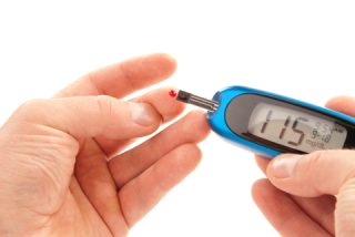
The long and short-term complications of uncontrolled diabetes can impact on a wide variety of parts of the body including eyes, heart, kidneys, nerves and feet. Read our 15 point checklist on keeping your feet healthy. To find out more about complications, and how to reduce the risk of developing them, read about managing wounds, loss of sensation (neuropathy) and loss of circulation (angiopathy). Other foot complications can include dry skin, clawing of toes and increased risk of stress fractures. With all complications, keeping blood glucose, blood pressure and blood fat levels under control will greatly help to reduce the risk of developing complications. Regular check-ups are essential to help manage the condition – find out what happens when you see the Podiatrist for your annual foot check here. World Diabetes Day – 14 November Eating healthily is the key message to managing both types of diabetes for this year’s World Diabetes Day (14 November every year). It is estimated that healthy eating could prevent up to 70% of Type 2 Diabetes. Check out the infographic below for quick advice on what to eat more or less of – just small changes every day for the whole family. Speak to your Podiatrist, Nutritionist and GP too about the steps you can take to improve your control. You can also find out more information online by searching for the ‘International Diabetes Federation’ and ‘Diabetes UAE’. Preventing Complications Preventing foot problems involves managing your diabetes well, controlling blood glucose levels (along with your cholesterol and blood pressure) and leading a healthy active lifestyle. Your chances of doing this will be great increased if you do not smoke. By adopting these measures, you can prevent or slow down any changes to the nerves and blood vessels that supply your legs and feet. In addition, regular foot care is important from preventing the build-up of hard skin to moisturizing your feet in order to keep the skin supple and always wearing the right shoes and socks that fit properly. Find out more about how to care for your feet here. Everyone who has diabetes should also have their feet checked regularly with a Podiatrist and at the very least once a year at their annual review. However, if you are at increased risk of complications, these checks may be done more frequently. When Should You See A Podiatrist? If you experience any form of neuropathy, pain or discomfort, it is important to consult your Diabetic Clinic or Podiatrist straight away, since it is easier to stop these developing into more serious issues such as ulcers or peripheral vascular disease. If you see any of the following in your feet, you should see your Podiatrist: • Walking becomes more difficult • Wearing shoes becomes more difficult • Tingling sensation / pins and needles • Part / all of your foot becomes swollen • Breaks in the skin, opens sores/blisters or a discharge • Skin colour changes (redder, bluer, paler, blacker) over part / all of the foot • Swelling in your feet and/or an unusual odour • Part / all of your foot feels hotter or colder than usual • Hard skin (callous) • Cramps in your calves • Shiny smooth skin and/or losing hair on your feet and legs Contact the leading UK qualified Podiatrists at Dubai Podiatry Centre, who are trained in the latest diabetic management and wound care through the UK’s NHS and private practice on +971 4 3435390.  

بقلم ميشيل شامبلين

في مدونتنا السابقة، نظرنا إلى الاختلافات بين مرض السكري من النوع 1 والنوع 2. دعونا نلقي نظرة على كيفية تأثير مرض السكري، إذا ترك دون علاج، على الصحة. والأهم من ذلك، التغييرات التي يمكننا جميعًا إجراؤها لتحسين صحتنا وتقليل خطر حدوث مضاعفات، بما في ذلك تناول الطعام الصحي.

التأثير على الصحة

يمكن أن تؤثر المضاعفات الطويلة والقصيرة المدى لمرض السكري غير المنضبط على مجموعة واسعة من أجزاء الجسم بما في ذلك العين والقلب والكلى والأعصاب والقدمين. اقرأ قائمتنا المرجعية المكونة من ١٥ نقطة حول الحفاظ على صحة قدميك. لمعرفة المزيد عن المضاعفات وكيفية تقليل خطر الإصابة بها، اقرأ عن إدارة الجروح وفقدان الإحساس (الاعتلال العصبي) وفقدان الدورة الدموية (اعتلال الأوعية الدموية). يمكن أن تشمل مضاعفات القدم الأخرى جفاف الجلد وتخلخل أصابع القدم وزيادة خطر الإصابة بكسور الإجهاد. مع جميع المضاعفات، فإن الحفاظ على مستويات السكر في الدم وضغط الدم والدهون في الدم تحت السيطرة سيساعد بشكل كبير على تقليل خطر حدوث مضاعفات. تعتبر الفحوصات المنتظمة ضرورية للمساعدة في إدارة الحالة – اكتشف ما يحدث عندما ترى طبيب الأقدام لإجراء فحص قدمك السنوي هنا.

اليوم العالمي لمرض السكري – ١٤ نوفمبر

إن تناول الطعام الصحي هو الرسالة الأساسية لإدارة كلا النوعين من مرض السكري في اليوم العالمي للسكري لهذا العام (١٤ نوفمبر من كل عام). وتشير التقديرات إلى أن الأكل الصحي يمكن أن يمنع ما يصل إلى ٧٠% من مرض السكري من النوع الثاني. قم بإلقاء نظرة على الرسم البياني أدناه للحصول على نصائح سريعة حول ما يجب تناوله أكثر أو أقل – فقط تغييرات صغيرة كل يوم لجميع أفراد الأسرة. تحدث إلى طبيب الأقدام وأخصائي التغذية والطبيب العام أيضًا حول الخطوات التي يمكنك اتخاذها لتحسين قدرتك على التحكم. يمكنك أيضًا معرفة المزيد من المعلومات عبر الإنترنت من خلال البحث عن “الاتحاد الدولي للسكري” و”مرض السكري في الإمارات العربية المتحدة”.

منع المضاعفات

تتضمن الوقاية من مشاكل القدم إدارة مرض السكري بشكل جيد، والتحكم في مستويات الجلوكوز في الدم (جنبًا إلى جنب مع الكوليسترول وضغط الدم) واتباع نمط حياة صحي نشط. ستزداد فرصك في القيام بذلك بشكل كبير إذا كنت لا تدخن. من خلال اعتماد هذه التدابير، يمكنك منع أو إبطاء أي تغييرات في الأعصاب والأوعية الدموية التي تغذي ساقيك وقدميك.

بالإضافة إلى ذلك، تعتبر العناية المنتظمة بالقدم أمرًا مهمًا بدءًا من منع تراكم الجلد القاسي إلى ترطيب قدميك من أجل الحفاظ على مرونة الجلد وارتداء الأحذية والجوارب المناسبة التي تناسبك دائمًا. تعرف على المزيد حول كيفية العناية بقدميك هنا.

يجب على كل شخص يعاني من مرض السكري أن يقوم بفحص قدميه بانتظام مع طبيب الأقدام وعلى الأقل مرة واحدة سنويًا في المراجعة السنوية. ومع ذلك، إذا كنت معرضًا لخطر متزايد لحدوث مضاعفات، فقد يتم إجراء هذه الفحوصات بشكل متكرر.

متى يجب أن ترى طبيب الأقدام؟

إذا كنت تعاني من أي شكل من أشكال الاعتلال العصبي أو الألم أو الانزعاج، فمن المهم استشارة عيادة مرضى السكري أو طبيب الأقدام على الفور، لأنه من الأسهل إيقاف تطورها إلى مشاكل أكثر خطورة مثل القرحة أو أمراض الأوعية الدموية الطرفية.

إذا رأيت أيًا مما يلي في قدميك، فيجب عليك مراجعة طبيب الأقدام الخاص بك:

يصبح المشي أكثر صعوبة

يصبح ارتداء الأحذية أكثر صعوبة

الإحساس بالوخز / الدبابيس والإبر

تورم جزء/كامل قدمك

حدوث تشققات في الجلد أو فتح القروح أو البثور أو الإفرازات

تغير لون الجلد (أحمر، أزرق، شاحب، أسود) على جزء/كل القدم

تورم في قدميك و/أو رائحة غير عادية

تشعر أن جزءًا من قدمك/كلها أكثر سخونة أو برودة من المعتاد

الجلد الصلب (القاسي)

تشنجات في ربلة الساق

بشرة ناعمة ولامعة و/أو تساقط الشعر في قدميك وساقيك

تواصل مع أبرز أطباء الأقدام المؤهلين في المملكة المتحدة في مركز دبي لعلاج الأرجل، والذين تم تدريبهم على أحدث علاجات مرض السكري والعناية بالجروح من خلال هيئة الخدمات الصحية الوطنية في المملكة المتحدة والعيادات الخاصة على الرقم ٠٤٣٤٣٥٣٩٠


Written by Michelle Champlin BSc Pod., M.Ch.S., S.R., Ch., (UK)

A bunion (hallux valgus) is a bony deformity at the base of the big toe, which can also cause the bone behind it, the metatarsal, to stick out. The big toe starts to point out towards the smaller toes. You can find out more about the increasing stages of bunions as they develop, what causes them and how to treat them.

Over-pronation is a significant factor in forefoot issues such bunionettes, neuromas and also in developing bunions, with many patients attending our clinic with bunions also exhibiting over-pronation of the sub-talar joints.

Over-pronation can lead to the collapse of your foot’s arches, leading to the appearance of flat feet, strain on the plantar fascia causing plantar fasciitis, as well as knee and hip problems.

Bunions, Tailors Bunions, Neuromas and Over-Pronation. When the sub-talar joint over-pronates and causes the ankle to roll in, this causes abnormal locking and unlocking of the midtarsal joint, resulting in the abnormal movement of the long bones in the forefoot called the metatarsal bones.

Bunions can sometimes be linked with overpronation

As this occurs, splaying of the metatarsals causes a widening of the forefoot and collapse of the transverse arch. Over time, this collapsed forefoot results in the formation of a bunion and / or a tailor’s bunion. The abnormal movement of the metatarsal bones can also result in the compression of the interdigital nerves in the ball of the foot causing painful neuromas. Neuromas in the forefoot are a swelling of the interdigital nerve as a result of chronic irritation from the abnormal movement of the metatarsal bones, which pinch the nerve. This causes swelling and damage to the nerve.

The abnormal locking and unlocking of the midtarsal joint also results in an imbalance between the tendons on the top of the toes and the tendons on the bottom of the toes. When this occurs, digital contracture results – or what are called hammertoes.

Treatment

Over-pronation and its resulting issues such as collapsed forefeet and bunions, are best treated early. There is no recommended home treatment other than the general avoidance of prolonged weight bearing in non-supportive footwear such as flip flops or ballet pumps until the patient can be assessed and treated by the Podiatrist. In the meantime, the area can be protected from irritation in footwear by wearing protective bunion gel sleeves or covers.

Treatment with custom-molded foot orthotics and regular footwear or sports shoes is critical to maintain stability of the foot and ankle and to improve and tighten the foot posture. The purpose of the orthotics is to get the subtalar joint functioning properly on its own again by tightening and toning the supporting ligaments, tendons and muscles, raising the transverse arch. Sometimes, if bunion formation is advanced, referral for specialist orthopaedic surgery may be advised.

If you notice that your foot seems to be getting wider (perhaps shoes beginning to pinch or cause irritation at your forefoot), pain in your feet or any other change in your foot’s posture, contact the expert Podiatrists at Dubai Podiatry Centre on +971 4 343590 | WhatsApp: +971 503553024.

بقلم ميشيل شامبلين

البروز العظمي هو تشوه عظمي في قاعدة إصبع القدم الكبير، والذي يمكن أن يتسبب أيضًا في بروز العظم خلفه، مشط القدم. يبدأ إصبع القدم الكبير في الإشارة نحو أصابع القدم الأصغر. يمكنك معرفة المزيد عن المراحل المتزايدة من الأورام مع تطورها، وأسبابها وكيفية علاجها.

يعد الكب الزائد عاملاً مهمًا في مشاكل مقدمة القدم مثل الأورام العصبية والأورام العصبية وأيضًا في تطور الأورام، حيث يعاني العديد من المرضى الذين يحضرون عيادتنا والذين يعانون من أورام يظهرون أيضًا فرط كب في المفاصل تحت الكاحل.

يمكن أن يؤدي الإفراط في الكب إلى انهيار أقواس قدمك، مما يؤدي إلى ظهور الأقدام المسطحة، والضغط على اللفافة الأخمصية مما يسبب التهاب اللفافة الأخمصية، بالإضافة إلى مشاكل في الركبة والورك.

الأورام، والأورام العصبية، والإفراط في الكب. عندما يفرط المفصل تحت الكاحل في الكب ويسبب انقلاب الكاحل للداخل، يؤدي ذلك إلى قفل وفتح غير طبيعي للمفصل الوسطي، مما يؤدي إلى حركة غير طبيعية للعظام الطويلة في مقدمة القدم والتي تسمى عظام مشط القدم.

في بعض الأحيان يمكن ربط الأورام بالكسر الزائد

عندما يحدث هذا، يؤدي تباعد مشط القدم إلى اتساع مقدمة القدم وانهيار القوس المستعرض. بمرور الوقت، يؤدي انهيار مقدمة القدم إلى تكوين ورم و/أو ورم الخياط. يمكن أن تؤدي الحركة غير الطبيعية لعظام مشط القدم أيضًا إلى ضغط الأعصاب بين الأصابع في كرة القدم مما يسبب ورمًا عصبيًا مؤلمًا. الأورام العصبية في مقدمة القدم هي تورم في العصب بين الأصابع نتيجة التهيج المزمن الناتج عن الحركة غير الطبيعية لعظام مشط القدم، مما يؤدي إلى قرص العصب. وهذا يسبب تورم وتلف العصب.

يؤدي القفل والفتح غير الطبيعي للمفصل الوسطي أيضًا إلى خلل في التوازن بين الأوتار الموجودة في الجزء العلوي من أصابع القدم والأوتار الموجودة في الجزء السفلي من أصابع القدم. عندما يحدث هذا، ينتج الانكماش الرقمي – أو ما يسمى أصابع المطرقة.

علاج

من الأفضل علاج الإفراط في الكب والمشاكل الناتجة عنه مثل انهيار القدم الأمامية والأورام في وقت مبكر. لا يوجد علاج منزلي موصى به بخلاف التجنب العام لتحمل الوزن لفترة طويلة بارتداء أحذية غير داعمة مثل الصنادل أو أحذية الباليه حتى يمكن تقييم المريض وعلاجه من قبل طبيب الأقدام. في هذه الأثناء، يمكن حماية المنطقة من التهيج بالأحذية من خلال ارتداء أكمام أو أغطية واقية من جل الورم.

يعد العلاج باستخدام أجهزة تقويم القدم المصممة خصيصًا والأحذية العادية أو الأحذية الرياضية أمرًا بالغ الأهمية للحفاظ على ثبات القدم والكاحل وتحسين وضعية القدم وتشديدها. الغرض من تقويم العظام هو جعل المفصل تحت الكاحل يعمل بشكل صحيح من تلقاء نفسه مرة أخرى عن طريق شد وتنعيم الأربطة والأوتار والعضلات الداعمة، مما يؤدي إلى رفع القوس المستعرض. في بعض الأحيان، إذا كان تكوين الورم متقدمًا، فقد يُنصح بالإحالة إلى جراحة العظام المتخصصة.

إذا لاحظت أن قدمك تبدو أكثر اتساعًا (ربما بدأ الحذاء يقرصك أو يسبب تهيجًا في مقدمة قدمك)، أو ألمًا في قدميك أو أي تغيير آخر في وضعية قدمك، اتصل بأطباء الأقدام الخبراء في مركز دبي للعناية بالاقدام على الرقم ٠٤٣٤٣٥٣٩٠ | واتساب: ٠٥٠٣٥٥٣٠٢٤


Written by Michelle Champlin BSc Pod., M.Ch.S., S.R., Ch., (UK) Infection can be caused by viruses, bacteria or fungus. In our foot clinic, the most common fungal infections seen is Athlete’s foot, an infection of the skin caused by a fungus (your feet can be home to over 100 types of funghi!). Plantar warts (verrucas) are caused by a viral infection of the skin. There are numerous types of bacterial infections – there are over ¼ million bacteria identified by microbiologists so far, of which about 400 can cause infection in humans. Infection generally requires a break in the skin from even a microscopic break in the skin from a cut, abrasion, puncture wound or ulcer. The most common infection is caused two bacteria, Staphylococcus and Streptococcus. Both of these infections cause tissue damage of various degrees. It is not uncommon to see a wound infected with various strains of different bacteria. Infection that occurs in the skin is called Cellulitis. Deep infections that develop puss pockets are called abscesses, like those in dentistry. The most common bacteria that causes Cellulitis is Streptococcus. These infections can become very serious and even life threatening. Cellulitis symptoms include: • spreading redness in the area • increase in the temperature of the skin • fever and chills. People who suffer from circulatory issues or chronic swelling in the legs (edema), are more at risk of developing these infections. Cellulitis is also known to be linked with athlete foot fungal conditions. The athlete’s foot causes tiny breaks in the skin, or even larger ‘fissures’ typically on the heel or between the toes, which can become infected by the Streptococcus bacteria. Bacteria that lives harmlessly on the surface of our skin can then cause infection by finding its way into our bodies and bloodstream through these tiny cuts. Soft corns, particularly between the toes, can also become infected and cause cellulitis and or an abscess. Puncture wounds, such as from animal bites, sea urchin wounds or standing on broken glass or nails, are very likely to become infected. This can result in a very dangerous deep abscess that can also infect the bone (called osteomyelitis). Your GP will prescribe antibiotics to fight the bacteria causing cellulitis A Doctor or Podiatrist should evaluate all deep puncture wounds as soon as possible. Simply cleaning the outside of the puncture wound is not enough to prevent infection as the bacteria may have been driven deep into the wound. Oral antibiotics are usually prescribed after thoroughly cleansing and dressing the wound, and the wound closely monitored. If there is any sign of infection, surgical cleaning of the wound is required. People with diabetes are at particular risk of infection. Diabetics spend more time in the hospital for foot infections than for any other reason. Corns and callouses on the feet of people with diabetes can break down and allow bacterial invasion of the tissue. In people with long standing open ulceration the underlying bone can become infected. Bone infections generally require surgery to remove the infected bone. These infections are very difficult to cure with oral or intra-venous antibiotics without also removing the infected bone. The presence of bone infection can be diagnosed with special tests such as bone scans, CT scans and MRI’s, in conjunction with the Doctor’s assessment. Whether Diabetic or not, this is why it is important to follow a regular foot care regime to keep your feet free of corns, to keep callous (hard skin) at a minimum and to moisturise daily with a specialist foot cream. This keeps skin healthy and free from cracks and less likely to allow bacteria, viruses or fungus to enter. Conditions such as gout can also lead to similar symptoms of redness and swelling, so it is important to see your Podiatrist or GP very early to obtain the correct diagnosis and treatment. Cellulitis requires urgent medical attention without delay. If you are worried about any pain, redness or swelling on your feet or legs, particularly after a trauma, contact Dubai Podiatry Centre immediately on +971 4 343590.  

بقلم ميشيل شامبلين

يمكن أن تحدث العدوى بسبب الفيروسات أو البكتيريا أو الفطريات. في عيادتنا للقدم، أكثر الالتهابات الفطرية شيوعًا هي قدم الرياضي، وهي عدوى جلدية تسببها الفطريات (يمكن أن تكون قدمك موطنًا لأكثر من 100 نوع من الفطريات!). الثآليل الأخمصية (verrucas) تنتج عن عدوى فيروسية في الجلد. هناك أنواع عديدة من الالتهابات البكتيرية – هناك أكثر من ربع مليون بكتيريا حددها علماء الأحياء الدقيقة حتى الآن، منها حوالي ٤٠٠ يمكن أن تسبب العدوى لدى البشر. تتطلب العدوى عمومًا حدوث كسر في الجلد حتى لو كان ذلك نتيجة لكسر مجهري في الجلد نتيجة لقطع أو تآكل أو جرح أو قرحة. العدوى الأكثر شيوعًا تسببها نوعان من البكتيريا، المكورات العنقودية والمكورات العقدية. كل من هذه العدوى تسبب تلف الأنسجة بدرجات مختلفة. ليس من غير المألوف رؤية جرح مصاب بسلالات مختلفة من البكتيريا المختلفة.

تسمى العدوى التي تحدث في الجلد بالتهاب النسيج الخلوي. تسمى الالتهابات العميقة التي تؤدي إلى ظهور جيوب قيحية بالخراجات، مثل تلك الموجودة في طب الأسنان. البكتيريا الأكثر شيوعًا التي تسبب التهاب النسيج الخلوي هي المكورات العقدية. يمكن أن تصبح هذه العدوى خطيرة للغاية وحتى مهددة للحياة.

تشمل أعراض التهاب النسيج الخلوي ما يلي:

انتشار الاحمرار في المنطقة

زيادة في درجة حرارة الجلد

الحمى والقشعريرة

الأشخاص الذين يعانون من مشاكل في الدورة الدموية أو تورم مزمن في الساقين (الوذمة)، هم أكثر عرضة للإصابة بهذه العدوى. ومن المعروف أيضًا أن التهاب النسيج الخلوي يرتبط بالحالات الفطرية للقدم الرياضي. تسبب قدم الرياضي شقوقًا صغيرة في الجلد، أو حتى “شقوقًا” أكبر عادةً على الكعب أو بين أصابع القدم، والتي يمكن أن تصاب بالعدوى بالبكتيريا العقدية. يمكن للبكتيريا التي تعيش على سطح بشرتنا بشكل غير ضار أن تسبب العدوى عن طريق إيجاد طريقها إلى أجسامنا ومجرى الدم من خلال هذه الجروح الصغيرة.

يمكن أيضًا أن تصاب مسامير القدم الناعمة، خاصة بين أصابع القدم، بالعدوى وتسبب التهاب النسيج الخلوي أو الخراج. من المحتمل جدًا أن تصاب بالعدوى الجروح الوخزية، مثل عضات الحيوانات أو جروح قنفذ البحر أو الوقوف على الزجاج المكسور أو الأظافر. يمكن أن يؤدي ذلك إلى خراج عميق خطير جدًا يمكن أن يصيب العظام أيضًا (يُسمى التهاب العظم والنقي).

سيصف طبيبك المضادات الحيوية لمحاربة البكتيريا المسببة لالتهاب النسيج الخلوي

يجب على الطبيب أو طبيب الأقدام تقييم جميع الجروح العميقة في أسرع وقت ممكن. إن مجرد تنظيف الجزء الخارجي من الجرح لا يكفي لمنع العدوى لأن البكتيريا ربما تكون قد تعمقت في الجرح. عادة ما يتم وصف المضادات الحيوية عن طريق الفم بعد تنظيف الجرح وتضميده جيدًا، ومراقبة الجرح عن كثب. إذا كان هناك أي علامة على العدوى، مطلوب التنظيف الجراحي للجرح.

الأشخاص المصابون بداء السكري معرضون بشكل خاص لخطر الإصابة. يقضي مرضى السكري وقتًا أطول في المستشفى بسبب التهابات القدم أكثر من أي سبب آخر. يمكن أن تتحلل مسامير القدم والثفن الموجودة على أقدام مرضى السكري وتسمح بغزو البكتيريا للأنسجة. في الأشخاص الذين يعانون من تقرح مفتوح طويل الأمد، يمكن أن يصاب العظم الأساسي بالعدوى. تتطلب التهابات العظام عمومًا إجراء عملية جراحية لإزالة العظام المصابة. يصعب علاج هذه العدوى بالمضادات الحيوية عن طريق الفم أو عن طريق الوريد دون إزالة العظم المصاب أيضًا. يمكن تشخيص وجود التهاب في العظام من خلال اختبارات خاصة مثل فحص العظام، والأشعة المقطعية، والرنين المغناطيسي، بالتزامن مع تقييم الطبيب.

سواء كنت مصابًا بداء السكري أم لا، لهذا السبب من المهم اتباع نظام منتظم للعناية بالقدمين للحفاظ على قدميك خاليتين من مسامير القدم، وللحفاظ على الجلد القاسي (الجلد الصلب) عند الحد الأدنى وترطيبهما يوميًا باستخدام كريم متخصص للقدم. وهذا يحافظ على صحة البشرة وخالية من التشققات ويقل احتمال السماح للبكتيريا أو الفيروسات أو الفطريات بالدخول.

يمكن أن تؤدي حالات مثل النقرس أيضًا إلى أعراض مشابهة من الاحمرار والتورم، لذلك من المهم رؤية طبيب الأقدام أو الطبيب العام في وقت مبكر جدًا للحصول على التشخيص والعلاج الصحيح. يتطلب التهاب النسيج الخلوي عناية طبية عاجلة دون تأخير.

إذا كنت قلقًا بشأن أي ألم أو احمرار أو تورم في قدميك أو ساقيك، خاصة بعد التعرض لصدمة، فاتصل بمركز دبي لعلاج الأقدام على الفور على الرقم ٠٤٣٤٣٥٣٩٠.


Written by Michelle Champlin BSc Pod., M.Ch.S., S.R., Ch., (UK) Flat feet is where the long arch on the inside of the foot is lowered or completely collapsed. Flat feet can develop in adulthood due to issues such as over-pronation, or be congenital – inherited and present since birth (pes planus). It is very important that children with flat feet are evaluated by a Podiatrist to determine if they need treatment to prevent future pain or deformity in their feet. This is one of the reasons why Dubai Podiatry Centre runs school health checks for children aged 5-6 years. When babies start to toddle at circa 9-15 months of age, the foot has a naturally chubby appearance as the bones and structures of the feet are still forming – this is perfectly normal. You can find out more about children’s foot development here. Arches in children’s feet usually develop by age 5 However, if the child still appears to have ‘flat feet’ with little or no noticeable arch at the age of about 5 years old, then it is wise to have the foot examined by a Podiatrist, advises Chief Podiatrist Michelle Champlin. Here are 5 signs to look out for in children that might signal a foot issue. Mrs Champlin explains that “The reason that it is important to check children’s feet at this age is because their little feet are still largely made of cartilage, with less bone than would be present in the adult foot. Since cartilage is relatively pliable, the abnormal forces caused by a flatfoot deformity may cause permanent structural alterations to the bones and joints of the foot that will continue and worsen into adulthood. This is the age where intervention, through the use of say orthotics in their school shoes, can prevent problems or pain in later life.” Symptoms Flat feet in children causes a number of changes to the structure of the foot which the Podiatrist will look for in a lower limb biomechanical assessment. Flat feet causes a number of musculoskeletal changes: the inside arch will be flattened, causing the heel bone (calcaneum) to be turned outward, and the inside of the foot to appear more bowed outward than normal, almost ‘banana’ shaped. Most cases of paediatric flat feet are associated with excessive flexibility in the joints of the foot which is commonly caused by ligamentous laxity – which Podiatrists call hypermobility. Since flat feet cause instability in the feet whilst walking, running or jumping, children with flat feet may have complaints in the foot such as arch, heel or ankle pain which is generally associated with increased standing, walking or running activities. The child may complain of feeling tired when walking round the mall for example, or not want to take part in sports and physical activities. They may also trip more than usual. Collapsed long arches in children leads to excessive rolling in of the arches of the foot, which also make the leg and knee turn inwards – children with flatfeet may also complain of pain in their lower back, hip, knee, or leg due to the misalignment of the foot and ankle. Treatment The good news is that flat feet in children (and adults) can be treated, usually with a slimline custom corrective orthotic that fits easily and discretely into their school and sports shoes. To have your children’s feet checked, contact the friendly Podiatrists at Dubai Podiatry Centre on +971 4 3435390.  

بقلم ميشيل شامبلين

القدم المسطحة هي المكان الذي ينخفض فيه القوس الطويل الموجود داخل القدم أو ينهار تمامًا. يمكن أن تتطور الأقدام المسطحة في مرحلة البلوغ بسبب مشاكل مثل الإفراط في الكب، أو أن تكون خلقية – موروثة وموجودة منذ الولادة (pes Planus). من المهم جدًا أن يتم تقييم الأطفال الذين يعانون من أقدام مسطحة من قبل طبيب الأقدام لتحديد ما إذا كانوا بحاجة إلى علاج لمنع الألم أو التشوه في أقدامهم في المستقبل. وهذا هو أحد الأسباب التي تدفع مركز دبي لعلاج الأرجل إلى إجراء فحوصات صحية مدرسية للأطفال الذين تتراوح أعمارهم بين ٥ و٦ سنوات.

عندما يبدأ الأطفال في الرضاعة في عمر ٩ إلى ١٥ شهرًا تقريبًا، يكون للقدم مظهر سمين بشكل طبيعي حيث لا تزال عظام وهياكل القدم في طور التكوين – وهذا أمر طبيعي تمامًا. يمكنك معرفة المزيد عن تطور قدم الأطفال هنا.

عادة ما تتطور الأقواس في أقدام الأطفال في سن الخامسة

ومع ذلك، إذا كان الطفل لا يزال يبدو وكأنه “أقدام مسطحة” مع القليل من القوس الملحوظ أو لا يوجد أي قوس ملحوظ عند عمر 5 سنوات تقريبًا، فمن الحكمة أن يتم فحص القدم من قبل طبيب الأقدام، كما تنصح رئيسة أطباء الأقدام ميشيل شامبلين. فيما يلي ٥ علامات يجب الانتباه إليها عند الأطفال والتي قد تشير إلى وجود مشكلة في القدم.

تشرح السيدة شامبلن أن “السبب وراء أهمية فحص أقدام الأطفال في هذا العمر هو أن أقدامهم الصغيرة لا تزال تتكون إلى حد كبير من الغضروف، مع كمية عظام أقل من تلك الموجودة في قدم البالغين. نظرًا لأن الغضروف مرن نسبيًا، فإن القوى غير الطبيعية الناتجة عن تشوه القدم المسطحة قد تسبب تغيرات هيكلية دائمة في عظام ومفاصل القدم والتي ستستمر وتتفاقم حتى مرحلة البلوغ. هذا هو العصر الذي يمكن فيه للتدخل، من خلال استخدام أجهزة تقويم العظام في أحذيتهم المدرسية، أن يمنع حدوث مشاكل أو ألم في وقت لاحق من الحياة.

أعراض

تسبب الأقدام المسطحة عند الأطفال عددًا من التغييرات في بنية القدم والتي سيبحث عنها طبيب الأقدام في التقييم الميكانيكي الحيوي للطرف السفلي. تسبب القدم المسطحة عددًا من التغيرات العضلية الهيكلية: يصبح القوس الداخلي مسطحًا، مما يتسبب في انحراف عظم الكعب (العقبي) إلى الخارج، ويظهر الجزء الداخلي من القدم أكثر انحناءًا إلى الخارج من الطبيعي، على شكل موزة تقريبًا.

ترتبط معظم حالات الأقدام المسطحة لدى الأطفال بالمرونة المفرطة في مفاصل القدم والتي تنتج عادةً عن تراخي الأربطة – وهو ما يطلق عليه أطباء الأقدام فرط الحركة.

نظرًا لأن القدم المسطحة تسبب عدم استقرار في القدم أثناء المشي أو الجري أو القفز، فقد يعاني الأطفال الذين يعانون من أقدام مسطحة من شكاوى في القدم مثل ألم القوس أو الكعب أو الكاحل الذي يرتبط عمومًا بزيادة أنشطة الوقوف أو المشي أو الجري. قد يشكو الطفل من الشعور بالتعب عند التجول في المركز التجاري على سبيل المثال، أو عدم الرغبة في المشاركة في الأنشطة الرياضية والبدنية. وقد يتعثرون أيضًا أكثر من المعتاد.

يؤدي انهيار الأقواس الطويلة عند الأطفال إلى التدحرج المفرط لأقواس القدم، مما يؤدي أيضًا إلى دوران الساق والركبة إلى الداخل – وقد يشتكي الأطفال الذين يعانون من الأقدام المسطحة أيضًا من ألم في أسفل الظهر أو الورك أو الركبة أو الساق بسبب اختلال المحاذاة. من القدم والكاحل.

علاج

والخبر السار هو أنه يمكن علاج الأقدام المسطحة لدى الأطفال (والكبار)، عادةً باستخدام جهاز تقويمي تصحيحي رفيع مخصص يتناسب بسهولة وبشكل منفصل مع أحذيتهم المدرسية والرياضية.

لفحص أقدام أطفالك، اتصل بأطباء الأقدام الودودين في مركز دبي لعلاج الأقدام على الرقم ٠٤٣٤٣٥٣٩٠


Written by Michelle Champlin BSc Pod., M.Ch.S., S.R., Ch., (UK)

Overlapping toes is a condition that can be present from birth (congenital) or more commonly, develop later on in life. The condition may affect the patient by being nothing more than unsightly and an inconvenience when selecting shoes, or in some cases cause extreme pain and discomfort when in shoes. Overlapping toes most commonly affect the big toe overlapping with the 2nd toe, or sometimes the little toe (5th) overlapping with the 4th.

Causes are varied – when the big toe is affected it can be linked to hallux valgus (bunion) which as it becomes worse causes the big toe to drift towards the 2nd toe. It can also be genetic.

Overlapping toes can be present from birth (congenital) or develop later in life


Treatment in Children

Effective conservative treatment depends upon how early the diagnosis is made. In young children, passive stretching and adhesive taping is most commonly used. This may require 6 to 12 weeks to accomplish, but reoccurrence is not uncommon. Chief Podiatrist Michelle Champlin urges all parents to bring their children to a Podiatrist for a foot check, to pick up issues such as overlapping toes or flat feet, as correction is generally much easier whilst the foot is still developing.


Treatment in Adults

Conservative treatment (non-surgical treatment) of overlapping toes starts with accommodating the overlapping toes. Shoes with a high, broad toe box are recommended and the Podiatrist will be able to advise in detail, with suggestions of brands that may be best for your individual foot shape. Forefoot supports such as gel toe straighteners and gel toe caps are often recommended to keep overlapping toes apart and prevent friction within footwear that can cause bursas or corns.

If the overlapping is linked with an underlying cause such as bunions, the Podiatrist can assess and advise corrective devices: slimline, discrete custom orthotics.

Rarely will the overlapping correct itself. As the individual matures the misalignment can become fixed. Sometimes, surgical correction by an orthopaedic surgeon specializing in feet may be suggested if the malalignment is fixed or severe, involving a skin plasty to release the contracture of the skin, a tendon release and a release of the soft tissues about the joint at the base of the affected toe may be required. In some cases the toe may require the placement of a pin to hold the toe in a straighten position. The pin, which exits the tip of the toe, may be left in place for up to three weeks. As with most foot surgery, there will be a period afterwards of immobility to maintain the surgeon’s work – with an aircast, splinting and possibly a post-operative orthotic to be worn within footwear.

For any toe, foot, ankle or leg issue contact the expert Podiatrists at Dubai Podiatry Centre Call: +971 4 3435390| WhatsApp: +971 50 3553024 | Book Online (click here)

بقلم ميشيل شامبلين

تداخل أصابع القدم هو حالة يمكن أن تكون موجودة منذ الولادة (خلقية) أو الأكثر شيوعًا أنها تتطور لاحقًا في الحياة. قد تؤثر الحالة على المريض من خلال كونها مجرد مظهر قبيح وإزعاج عند اختيار الأحذية، أو في بعض الحالات تسبب ألمًا شديدًا وعدم راحة عند ارتداء الأحذية. تؤثر أصابع القدم المتداخلة بشكل شائع على تداخل إصبع القدم الكبير مع إصبع القدم الثاني، أو في بعض الأحيان تداخل إصبع القدم الصغير (الخامس) مع الإصبع الرابع.

تتنوع الأسباب – عندما يتأثر إصبع القدم الكبير، يمكن أن يكون مرتبطًا بإبهام القدم الأروح (الوكعة) والذي عندما يصبح أسوأ يؤدي إلى انجراف إصبع القدم الكبير نحو إصبع القدم الثاني. ويمكن أيضا أن تكون وراثية.

يمكن أن تكون أصابع القدم المتداخلة موجودة منذ الولادة (خلقية) أو تتطور لاحقًا في الحياة

العلاج عند الأطفال

يعتمد العلاج المحافظ الفعال على مدى التشخيص المبكر. عند الأطفال الصغار، يتم استخدام التمدد السلبي والشريط اللاصق بشكل شائع. قد يتطلب ذلك من 6 إلى 12 أسبوعًا لإنجازه، ولكن تكرار ذلك ليس أمرًا غير شائع. تحث رئيسة أطباء الأقدام ميشيل شامبلين جميع الآباء على إحضار أطفالهم إلى أخصائي أمراض الأقدام لفحص القدم، لاكتشاف مشكلات مثل تداخل أصابع القدم أو القدم المسطحة، حيث يكون التصحيح أسهل بكثير بشكل عام بينما لا تزال القدم في طور النمو

العلاج عند البالغين

يبدأ العلاج المحافظ (العلاج غير الجراحي) لتداخل أصابع القدم باستيعاب أصابع القدم المتداخلة. يوصى بارتداء أحذية ذات مقدمة مرتفعة وعريضة، وسيتمكن طبيب الأقدام من تقديم المشورة بالتفصيل، مع اقتراحات للعلامات التجارية التي قد تكون الأفضل لشكل قدمك الفردي. غالبًا ما يُنصح باستخدام دعامات مقدمة القدم مثل أدوات فرد أصابع القدم الهلامية وأغطية أصابع القدم الهلامية للحفاظ على تداخل أصابع القدم بعيدًا ومنع الاحتكاك داخل الأحذية الذي يمكن أن يسبب الجراب أو مسامير القدم.

إذا كان التداخل مرتبطًا لسبب كامن مثل الأورام، فيمكن لطبيب الأقدام تقييم الأجهزة التصحيحية وتقديم المشورة لها: أجهزة تقويم العظام المخصصة الرفيعة والمنفصلة.

نادرًا ما يقوم التداخل بتصحيح نفسه. عندما ينضج الفرد، يمكن أن يصبح الاختلال ثابتًا. في بعض الأحيان، قد يتم اقتراح التصحيح الجراحي بواسطة جراح عظام متخصص في القدمين إذا كان سوء الاستقامة ثابتًا أو شديدًا، بما في ذلك رأب الجلد لتحرير تقلص الجلد، وإطلاق الوتر، وإطلاق الأنسجة الرخوة حول المفصل في القاعدة. قد تكون هناك حاجة لإصبع القدم المتضررة. في بعض الحالات، قد يتطلب إصبع القدم وضع دبوس لتثبيت إصبع القدم في وضع مستقيم. يمكن ترك الدبوس الذي يخرج من طرف إصبع القدم في مكانه لمدة تصل إلى ثلاثة أسابيع. كما هو الحال مع معظم جراحات القدم، ستكون هناك فترة بعد ذلك من عدم القدرة على الحركة للحفاظ على عمل الجراح – مع استخدام جبيرة هوائية، وربما استخدام جهاز تقويم العظام بعد العملية الجراحية داخل الأحذية.

في حالة وجود أي مشكلة في أصابع القدم أو القدم أو الكاحل أو الساق، اتصل بأخصائيي الأقدام الخبراء في مركز دبي لعلاج الأرجل اتصل على:043435390 | واتساب: 0503553024  |


Copyright Dubai Podiatry 2026. All rights reserved.

WhatsApp us