fbpx

Please wait...


BLOG

No more posts

Written by Michelle Champlin BSc Pod., M.Ch.S., S.R., Ch., (UK)
Underlapping toes are commonly seen in both adults and children. The toes most often involved are the fourth and fifth toes. A special form of underlapping toes is called ‘clinodactyly’ or congenital curly toes. Clinodactyly is fairly common and is inherited. One or more toes can be involved and toes three, four, and five of both feet are most commonly affected. For more information on overlapping toes, read more here.
The cause of the condition is not proven. One possible cause is an imbalance in muscle strength of the small muscles of the foot. This is aggravated by a subtle abnormality in the orientation on the joints in the foot just below the important ankle joint called the subtalar joint. This results in an abnormal pull on the ligaments in the toes causing them to curl. Then with weight bearing the malalignment is increased and a folding or curling of the toes results in the formation of callous on the outside margin of the end of the toe. Tight fitting shoes can aggravate the condition, causing blisters, bursas or corns on any areas that rub.
Treatment
The age of the patient, how underlapped the toes are and symptoms determine course of treatment. When intervention is suggested, the degree of underlapping determines the level of correction. When the condition is flexible a simple release of the tendon in the bottom of the toe will allow for straightening of the toe. If it is is rigid in nature then removal of a small portion of the bone in the toe may be necessary. Both of these procedures are common in adult patients for the correction of hammertoe deformity. If skin contracture is present a skin plasty may also be required.
The toes can also be protected from in-shoe friction by using gel lined protective toe caps or sleeves.
Toe alignment issues, especially linked to underlying foot conditions such as pes cavus (high arches) or bunions may also be addressed with the use of in-shoe custom orthotics.
For toe or foot malalignments, it is best to seek assessment early from a Podiatrist. Chief Podiatrist Michelle Champlin advises all children starting school to have their feet checked at about age 5, whilst the bones are still forming and any treatment can best take effect.
If you are concerned about your own or your children’s feet, contact Dubai Podiatry Centre on +971 4 3435390.


بقلم ميشيل شامبلين

تظهر أصابع القدم السفلية بشكل شائع عند البالغين والأطفال. أصابع القدم الأكثر إصابة هي أصابع القدم الرابعة والخامسة. يُطلق على شكل خاص من أصابع القدم المتداخلة اسم “أصابع القدم المتعرجة” أو أصابع القدم المتعرجة الخلقية. يعتبر مرض الإكلينوداكتيل شائعًا إلى حد ما وهو موروث. يمكن أن يصاب إصبع واحد أو أكثر من أصابع القدم، وتكون أصابع القدم الثلاثة والأربعة والخمسة هي الأكثر شيوعًا. لمزيد من المعلومات حول تداخل أصابع القدم، اقرأ المزيد هنا.

لم يتم إثبات سبب هذه الحالة. أحد الأسباب المحتملة هو خلل في قوة العضلات في العضلات الصغيرة للقدم. ويتفاقم هذا بسبب وجود خلل طفيف في الاتجاه على المفاصل في القدم أسفل مفصل الكاحل المهم الذي يسمى المفصل تحت الكاحل. وينتج عن هذا سحب غير طبيعي على أربطة أصابع القدم مما يؤدي إلى تجعدها. ثم مع تحمل الوزن يزداد سوء المحاذاة ويؤدي طي أو تجعيد أصابع القدم إلى تكوين قاسية على الحافة الخارجية لنهاية إصبع القدم. يمكن أن تؤدي الأحذية الضيقة إلى تفاقم الحالة، مما يسبب ظهور بثور أو أكياس أو مسامير في أي منطقة تحتك.

العلاج: عمر المريض، ومدى تداخل أصابع القدم، والأعراض هي التي تحدد مسار العلاج. عندما يقترح التدخل، فإن درجة التداخل تحدد مستوى التصحيح. عندما تكون الحالة مرنة، فإن التحرير البسيط للوتر الموجود في الجزء السفلي من إصبع القدم سيسمح بتقويم إصبع القدم. إذا كانت صلبة بطبيعتها، فقد يكون من الضروري إزالة جزء صغير من العظم الموجود في إصبع القدم. كلا الإجراءين شائعان لدى المرضى البالغين لتصحيح تشوه إصبع القدم المطرقية. في حالة وجود تقلص الجلد، قد تكون هناك حاجة أيضًا إلى رأب الجلد.

يمكن أيضًا حماية أصابع القدم من الاحتكاك داخل الحذاء باستخدام أغطية أو أكمام واقية لأصابع القدم مبطنة بالجيل.

يمكن أيضًا معالجة مشكلات محاذاة إصبع القدم، وخاصة المرتبطة بحالات القدم الأساسية مثل pes cavus (الأقواس العالية) أو الأورام باستخدام أجهزة تقويم العظام المخصصة داخل الحذاء.

بالنسبة لتشوهات أصابع القدم أو القدم، فمن الأفضل طلب التقييم مبكرًا من طبيب الأقدام. تنصح رئيسة أطباء الأقدام ميشيل شامبلين جميع الأطفال الذين يبدأون المدرسة بفحص أقدامهم عند سن الخامسة تقريبًا، بينما لا تزال العظام في طور التشكل وأي علاج يمكن أن يكون له تأثير أفضل.

إذا كنت قلقًا بشأن قدمك أو قدم أطفالك، اتصل بمركز دبي لعلاج الأرجل على الرقم ٠٤٣٤٣٥٣٩٠


Insoles

Written by Michelle Champlin BSc Pod., M.Ch.S., S.R., Ch., (UK)
Many people require custom orthotics for a range of reasons:
• foot and leg issues (fallen arches, high arches, aching, tired feet, heel pain, pain in the ball of the foot, corns, calluses, overpronation, bunions)
• Sports injuries (Achilles tendon pain, shin splints, knee pain, hip or back pain)
• Musculoskeletal problems (back pain, myalgia, fibrositis, arthritis)
• Diabetes, e.g. prevention of foot ulcers by offloading high pressure areas

From boaters to firemen, lots of patients need water resistant custom orthotics
Lots of these biomechanics patients that we treat further require ‘waterproof’ custom orthotics due to either their recreational pursuits or type of work they do. Anyone from kayakers, yacht/motorboat sailors, firemen or PE teachers can spend many hours in wet environments. Thus, many of these patients’ feet are exposed to high levels of moisture – and others just perspire a lot (especially in our hot climate) in their work or school shoes throughout the day. Exposure to moisture and perspiration can cause materials such as leather or foam, commonly used to make some insoles, to crack and degrade quickly.
At Dubai Podiatry Centre, our Podiatrists hand make all of our patients fully custom orthotics from a mold of the patients’ feet in our in-clinic orthotics laboratory. For orthotics requiring water resistance, we tend to mill the orthotics from a high medical grade PPP resin. This device is completely waterproof and dries off in seconds. For a cover / cushioning, neoprene type medical grade material is used, in a variety of milimetre thicknesses depending on prescription. This material is antibacterial and antifungal. Sometimes, a special shock absorbing medical material called poron can also be integrated. Even with extremely heavy use, the ‘shell’ of the orthotic will stay the same and the orthotic covering can just be refurbished as and when required. Because our fully equipped lab is in-clinic and we don’t send away to factories for generic manufacture, we can normally carry out adjustments or refurbishments on the same day or whilst you wait!
To keep your orthotics as fresh as the day you got them from us, we recommend wiping them down with a little antibacterial / antifungal essential oil such as lavender oil, tea tree oil or Japanese Peppermint.
For thorough investigation of any foot or leg issue and expert orthotic therapy, contact Dubai Podiatry Centre on +971 4 3435390. Based on Sheikh Zayed Road, Dubai for over 15 years, the clinic is led by Chief Podiatrist Michelle Champlin. Conveniently located opposite the DIFC district, the Podiatrists are UK university trained and qualified and extensively experienced in both NHS and private clinic practice.


بقلم ميشيل شامبلين

يحتاج العديد من الأشخاص إلى أجهزة تقويم العظام المخصصة لمجموعة من الأسباب:

مشاكل في القدم والساق (سقوط الأقواس، والأقواس المرتفعة، والألم، والقدم المتعبة، وألم الكعب، وألم في مشط القدم، والذرة، والدُشبذات، والكَبّ الزائد، والأورام)

الإصابات الرياضية (ألم في وتر العرقوب، وشظايا قصبة الساق، وألم في الركبة، وألم في الورك أو الظهر)

مشاكل العضلات والعظام (آلام الظهر، وألم عضلي، والتهاب الليفي، والتهاب المفاصل)

مرض السكري، على سبيل المثال. الوقاية من تقرحات القدم عن طريق تفريغ مناطق الضغط المرتفع

من رواد القوارب إلى رجال الإطفاء، يحتاج الكثير من المرضى إلى أجهزة تقويم العظام المخصصة المقاومة للماء

يحتاج الكثير من مرضى الميكانيكا الحيوية الذين نعالجهم أيضًا إلى أجهزة تقويم العظام المخصصة “المقاومة للماء” إما بسبب مساعيهم الترفيهية أو نوع العمل الذي يقومون به. يمكن لأي شخص من زوارق الكاياك أو بحارة اليخوت/القوارب البخارية أو رجال الإطفاء أو معلمي التربية البدنية قضاء ساعات طويلة في البيئات الرطبة. وبالتالي، تتعرض أقدام العديد من هؤلاء المرضى لمستويات عالية من الرطوبة – والبعض الآخر يتعرق كثيرًا (خاصة في مناخنا الحار) في أحذية العمل أو المدرسة طوال اليوم. يمكن أن يؤدي التعرض للرطوبة والعرق إلى تشقق وتحلل مواد مثل الجلد أو الرغوة، التي تستخدم عادة لصنع بعض النعال الداخلية.

في مركز دبي لطب الأقدام، يقوم أطباء الأقدام لدينا بتصنيع أجهزة تقويم العظام المخصصة لجميع مرضانا من قالب أقدام المرضى في مختبر تقويم العظام داخل العيادة. بالنسبة إلى أجهزة تقويم العظام التي تتطلب مقاومة للماء، فإننا نميل إلى طحن أجهزة تقويم العظام من راتنجات PPP عالية الجودة من الدرجة الطبية. هذا الجهاز مقاوم للماء تمامًا ويجف في ثوانٍ. بالنسبة للغطاء/الوسادة، يتم استخدام مادة طبية من نوع النيوبرين بسماكات مختلفة حسب الوصفة الطبية. هذه المادة مضادة للبكتيريا والفطريات. في بعض الأحيان، يمكن أيضًا دمج مادة طبية خاصة ممتصة للصدمات تسمى البورون. حتى مع الاستخدام المكثف للغاية، ستظل “غلاف” جهاز تقويم العظام كما هو ويمكن تجديد غطاء تقويم العظام عند الحاجة. نظرًا لأن مختبرنا المجهز بالكامل موجود في العيادة ولا نرسله بعيدًا إلى المصانع لتصنيع الأدوية العامة، فيمكننا عادةً إجراء التعديلات أو التجديدات في نفس اليوم أو أثناء الانتظار!

للحفاظ على تقويم العظام الخاص بك طازجًا كما هو الحال في اليوم الذي حصلت فيه عليه منا، نوصي بمسحه باستخدام القليل من الزيت العطري المضاد للبكتيريا / المضاد للفطريات مثل زيت اللافندر أو زيت شجرة الشاي أو النعناع الياباني.

لإجراء فحص شامل لأي مشكلة في القدم أو الساق وعلاج تقويم العظام على يد خبراء، اتصل بمركزمركز دبي للعناية بالاقدام على الرقم 043435390 . تقع العيادة في شارع الشيخ زايد، دبي منذ أكثر من 15 عامًا، وتديرها رئيسة أطباء الأقدام ميشيل شامبلين. يتمتع أطباء الأقدام بموقع ملائم مقابل منطقة مركز دبي المالي العالمي، وهم مدربون ومؤهلون من جامعة المملكة المتحدة ويتمتعون بخبرة واسعة في كل من الخدمات الصحية الوطنية والعيادات الخاصة.


Written by Michelle Champlin BSc Pod., M.Ch.S., S.R., Ch., (UK)
Knee pain in children can have a number of causes. Knee pain, as with any heel, foot or leg pain in children should be evaluated by your doctor or Podiatrist. Dubai Podiatry Centre specializes in biomechanics and ‘podo-paediatrics’ – the foot and leg healthcare of children.
Osgood-Schlatter’s Disease
A common cause of knee pain in a growing child is injury to a growth plate at the front of the knee. This may be accompanied by swelling in the area. This is called Osgood-Schlatter’s disease. It occurs most commonly in children between the ages of 10 -14. It is most often associated with very active children and cycling. It is thought that the condition is mainly caused by an abnormal pull of the tendon that attaches into the painful area from the kneecap – the patellar tendon. Read more about Osgood-Schlatter’s here.

Sporty children are at higher risk of developing Osgood-Schlatter’s Disease
Treatment
Treatment starts with avoiding activities that aggravate the condition such as kneeling or excessive flexion of the knee. Icing the area and anti-inflammatory medications (NSAIDs) for pain relief. Often the abnormal knee function is due to the way the foot functions. In this instance the foot causes the lower leg to rotate slightly inward. This causes the patellar tendon to pull in an abnormal direction where it attaches in front of the knee causing damage to the growth plate in this area. If this is the cause of the problem then treatment will usually involve wearing custom corrective orthotics. Dubai Podiatry Centre’s Podiatrists take a mold of the child’s feet, then make the orthotics in their laboratory in the clinic. These are then fitted into the child’s school and/or sports shoes. The orthotic corrects the underlying foot condition that is affecting the lower leg and knee. As with any lower limb biomechanics issues, especially those involving the developing feet of children, it is important that a Podiatrist trained in the use of orthotics should be able to evaluate the cause of the problem and treat it properly.

If your child is complaining of any leg, foot or knee pain, contact the experts at Dubai Podiatry Centre on +971 4 3435390.

بقلم ميشيل شامبلين

يمكن أن يكون لألم الركبة عند الأطفال عدد من الأسباب. يجب تقييم آلام الركبة، كما هو الحال مع أي ألم في الكعب أو القدم أو الساق عند الأطفال، من قبل طبيبك أو طبيب الأقدام. يتخصص مركز دبي لطب الأقدام في الميكانيكا الحيوية و”طب أقدام الأطفال” – الرعاية الصحية للقدم والساق لدى الأطفال.

مرض أوسجود-شلاتر

أحد الأسباب الشائعة لألم الركبة عند الأطفال أثناء نموهم هو إصابة لوحة النمو في الجزء الأمامي من الركبة. وقد يصاحب ذلك تورم في المنطقة. وهذا ما يسمى مرض أوسجود شلاتر. ويحدث بشكل شائع عند الأطفال الذين تتراوح أعمارهم بين 10 إلى 14 عامًا. غالبًا ما يرتبط بالأطفال النشطين جدًا وركوب الدراجات. يُعتقد أن هذه الحالة تنجم بشكل أساسي عن سحب غير طبيعي للوتر الذي يرتبط بالمنطقة المؤلمة من الرضفة – الوتر الرضفي. اقرأ المزيد عن Osgood-Schlatter هنا.

الأطفال الرياضيون أكثر عرضة للإصابة بمرض أوسجود-شلاتر

علاج

يبدأ العلاج بتجنب الأنشطة التي تؤدي إلى تفاقم الحالة مثل الركوع أو الثني المفرط للركبة. وضع الثلج على المنطقة والأدوية المضادة للالتهابات (NSAIDs) لتخفيف الألم. في كثير من الأحيان تكون وظيفة الركبة غير الطبيعية بسبب الطريقة التي تعمل بها القدم. في هذه الحالة، تتسبب القدم في دوران الجزء السفلي من الساق إلى الداخل قليلاً. يؤدي هذا إلى سحب الوتر الرضفي في اتجاه غير طبيعي حيث يعلق أمام الركبة مما يتسبب في تلف لوحة النمو في هذه المنطقة. إذا كان هذا هو سبب المشكلة، فإن العلاج عادةً ما يتضمن ارتداء أجهزة تقويم العظام التصحيحية المخصصة. يقوم أطباء الأقدام في مركز دبي لعلاج الأرجل بأخذ قالب لقدم الطفل، ثم يصنعون أجهزة تقويم العظام في مختبرهم في العيادة. ويتم بعد ذلك تركيبها في أحذية المدرسة و/أو الأحذية الرياضية للطفل. يقوم جهاز تقويم العظام بتصحيح حالة القدم الأساسية التي تؤثر على الجزء السفلي من الساق والركبة. كما هو الحال مع أي مشكلات تتعلق بالميكانيكا الحيوية في الأطراف السفلية، خاصة تلك التي تتعلق بالأقدام النامية لدى الأطفال، من المهم أن يكون طبيب الأقدام المدرب على استخدام تقويم العظام قادرًا على تقييم سبب المشكلة وعلاجها بشكل صحيح.

إذا كان طفلك يشكو من أي ألم في الساق أو القدم أو الركبة، تواصل مع الخبراء في مركز دبي

043435390 للعناية بالاقدام على


One of the fastest growing sports in the Middle East is the triathlon. Both amateur and professional events happen almost every weekend in the region, including Tri Yas and Al Ain Multisports (outside of summer months). Triathlon involves swimming, biking and running, with little or no rest in between.

Race distances vary and finish times range from approximately 30 minutes to several days as athletes compete in swimming, cycling and running. Although the Ironman distance race is probably the most well known, races of Olympic distance or shorter are the most popular.
The most common triathlon injuries that are seen at Dubai Podiatry Centre, according to Chief Podiatrist Michelle Champlin, are injuries of the leg below the knee. Frequent acute injuries include bruises, abrasions, lacerations, blisters, sprains, strains and fractures. More chronic injuries include runners knee, ITB syndrome or shin splints.
Acute Injuries
Bruising/contusions: usually caused by collisions or falls. These injuries can result in hemorrhage. Blood vessels under the skin in the tissues usually tear and varying amounts of blood and plasma leak into the wound, and produce swelling and pain.
Small bruises require no special emergency care. Rest, ice, compression and elevation, along with stretching and range of motion exercises, are usually all that is needed. With severe closed soft tissue injuries, extensive swelling and bleeding beneath the skin may occur. Apply a padding and a soft roller bandage for pressure, which can limit this bleeding. Elevating the affected leg and applying ice to the area can be helpful in decreasing bleeding of injured tissue and reducing tissue swelling. If the athlete has suffered extensive soft tissue damage, see a Podiatrist or Doctor for investigation re a possible bone fracture as well.
Blisters: Foot blisters are among the most common injuries for athletes, caused by friction. Small, intact blisters that don’t cause discomfort usually don’t need treatment. The best protection against infection is a blister’s own skin roof. To protect the roof, you can cover this type of blister with a small adhesive bandage. You should drain larger or painful blisters without removing the roof – you can find out more about lancing blisters here. The best action is of course prevention – find out more about buying running shoes here, and our Podiatrist’s specific ‘save-a-trek’ blister prevention packs to treat hot spots. See your Podiatrist for larger or deep blisters for treatment and appropriate topical medications / dressing.
Abrasions: Most often caused by a crash or fall from the bicycle or by a runner losing their footing and falling on a rough surface, such as Wadi Bih. The skin is abraded and can become a deep wound, with embedded dirt. First-degree injuries are superficial, second-degree injuries leave some or partial thickness damage to the skin and third degree is full-thickness damage to abraded areas.
Treatment: clean it out very thoroughly, removing all debris (i.e., gravel, sand or dirt) from the skin tissue. You may use betadine soap or similar and gauze to scrub the area clean. If debris is embedded deep in the tissue, see your Podiatrist or GP immediately, who can ensure the wound is deeply cleaned and sterile. Once it is clean, cover it with a non-stick sterile dressing. Second-degree abrasions and wounds normally respond to the same care as first-degree wounds. Third-degree wounds require medical attention with skin grafting occasionally necessary.
Lacerations: These injuries commonly occur from falling off bicycles or cuts from rocks during the swim stage. Common sites for lacerations at a triathlon include the hands, arms, legs, feet and head.
Treatment: control bleeding from the wound with direct pressure. After bleeding is under control, clean the wound with soap and water. Then apply an antiseptic solution such as Betadine. Close the wound by pulling the edges of the wound together with adhesive strips such as Steri-Strips. Assess larger lacerations and see a Doctor if stitches are required. It is important to remember all open wounds should be kept clean and the bandages changed regularly. Observe wounds for signs of infection (swelling, redness, pus etc.).
Muscle strains: These are a result of a sudden muscle overload, causing the fibres of the muscle and tendon to tear. The signs and symptoms of muscle strain include pain, muscle spasm and loss of strength.
Treatment: Athletes should rest, ice, compress and elevate. This reduces tissue damage from bleeding and swelling. Take extra caution when placing ice directly over nerves, such as on the top of the foot, to prevent damage from cold exposure. Strains that are too painful for normal walking or that cause pain when the athlete moves the injured part should be protected and rested, and a Podiatrist consulted. Read our 15 tips for avoiding running-related injuries here for the run stage.
Ligament sprains: Ligament sprains, especially of the ankle, are common in endurance races such as triathlons, particularly in runners on rough or unstable (i.e. beach) terrain. The management of a ligament sprain depends on the degree of injury.
Treatment: rest, ice, compression and elevation initially. See a Podiatrist for all strains, as these can be potentially as serious as fractures in terms of consequences for your biomechanics. Serious strains may require using an ‘aircast’ to immobilize the ankle. The Podiatrist will provide a recovery treatment plan, including exercises and identify any underlying cause of the injury. Completely torn ligaments may require surgery.
Fractures: Acute fractures are common in the cycling stage, and may be open or closed. A good indication that there may be a fracture is the athlete’s inability to use the extremity, extreme pain and swelling. An athlete with a fracture or serious injury usually “guards” the injured part and refuses to use it because motion or weight bearing increases pain. Occasionally, non-displaced fractures are not very painful and many athletes, especially those participating in triathlons, will continue to use a painful limb and “run through” the pain.
Treatment: The management of fractures on-site is similar to that of treating severe sprains and includes ice, elevation, compression and immobilization. Cover any open wounds with a dry, non-linty sterile dressing and apply pressure to control bleeding. Splint a fracture before moving the athlete, and attend hospital immediately for emergency care.
For either acute or chronic foot, leg or hip injuries, contact the UK trained Podiatrists at Dubai Podiatry Centre on +971 4 3435390.


واحدة من أسرع الرياضات نموًا في الشرق الأوسط هي رياضة الترياتلون. تقام فعاليات الهواة والمحترفين تقريبًا في نهاية كل أسبوع في المنطقة، بما في ذلك Tri Yas وAl Ain Multisports (خارج أشهر الصيف). يتضمن الترياتلون السباحة وركوب الدراجات والجري، مع القليل من الراحة أو عدم الراحة بينهما.

تختلف مسافات السباق وتتراوح أوقات الانتهاء من حوالي 30 دقيقة إلى عدة أيام حيث يتنافس الرياضيون في السباحة وركوب الدراجات والجري. على الرغم من أن سباق مسافة الرجل الحديدي ربما يكون الأكثر شهرة، إلا أن سباقات المسافات الأولمبية أو الأقصر هي الأكثر شعبية.

إصابات الترياتلون الأكثر شيوعًا التي يتم مشاهدتها في مركز دبي لعلاج الأرجل، وفقًا لرئيس أطباء الأقدام ميشيل شامبلين، هي إصابات الساق تحت الركبة. وتشمل الإصابات الحادة المتكررة الكدمات والسحجات والتمزقات والبثور والالتواء والسلالات والكسور. المزيد من الإصابات المزمنة تشمل ركبة العدائين، أو متلازمة ITB، أو جبائر قصبة الساق.

الإصابات الحادة

الكدمات/الكدمات: عادة ما تكون ناجمة عن الاصطدامات أو السقوط. هذه الإصابات يمكن أن تؤدي إلى النزيف. عادةً ما تتمزق الأوعية الدموية الموجودة تحت الجلد في الأنسجة وتتسرب كميات متفاوتة من الدم والبلازما إلى الجرح، مما يسبب التورم والألم.

لا تتطلب الكدمات الصغيرة رعاية طارئة خاصة. كل ما هو مطلوب عادة هو الراحة والثلج والضغط والارتفاع، إلى جانب تمارين التمدد ونطاق الحركة. مع إصابات الأنسجة الرخوة الشديدة المغلقة، قد يحدث تورم ونزيف واسع النطاق تحت الجلد. ضع حشوة وضمادة دوارة ناعمة للضغط، مما قد يحد من هذا النزيف. يمكن أن يكون رفع الساق المصابة ووضع الثلج على المنطقة مفيدًا في تقليل نزيف الأنسجة المصابة وتقليل تورم الأنسجة. إذا عانى الرياضي من تلف شديد في الأنسجة الرخوة، قم بزيارة طبيب الأقدام أو الطبيب للتحقيق في احتمال وجود كسر في العظام أيضًا.

البثور: تعتبر بثور القدم من أكثر الإصابات شيوعًا بين الرياضيين، والتي تحدث بسبب الاحتكاك. البثور الصغيرة السليمة التي لا تسبب الانزعاج لا تحتاج عادة إلى علاج. أفضل حماية ضد العدوى هي تغطية سطح الجلد الخاص بالبثور. لحماية السقف، يمكنك تغطية هذا النوع من البثرة بضمادة لاصقة صغيرة. يجب عليك تصريف البثور الأكبر حجمًا أو المؤلمة دون إزالة السقف – يمكنك معرفة المزيد عن وخز البثور هنا. أفضل إجراء هو الوقاية بالطبع – اكتشف المزيد حول شراء أحذية الجري هنا، وحزم الوقاية من البثور الخاصة بأخصائيي الأقدام لدينا لعلاج النقاط الساخنة. راجع طبيب الأقدام الخاص بك للحصول على بثور أكبر أو عميقة للعلاج والأدوية / الضمادات الموضعية المناسبة.

السحجات: تحدث غالبًا بسبب اصطدام أو سقوط من دراجة هوائية أو بسبب فقدان العداء لقدمه وسقوطه على سطح خشن، مثل وادي بيه. يتآكل الجلد ويمكن أن يصبح جرحًا عميقًا مع أوساخ مدمجة. إصابات الدرجة الأولى سطحية، وإصابات الدرجة الثانية تترك ضررًا جزئيًا أو جزئيًا على الجلد، والدرجة الثالثة هي أضرار كاملة السُمك للمناطق المتآكلة. العلاج: تنظيفه جيدًا، وإزالة جميع الحطام (أي الحصى والرمل والأوساخ) من أنسجة الجلد. يمكنك استخدام صابون البيتادين أو ما شابه ذلك والشاش لفرك المنطقة وتنظيفها. إذا كانت الحطام مغروسة بعمق في الأنسجة، فراجع طبيب الأقدام أو الطبيب العام على الفور، والذي يمكنه التأكد من تنظيف الجرح بعمق وتعقيمه. بمجرد تنظيفه، قم بتغطيته بضمادة معقمة غير لاصقة. تستجيب الجروح والسحجات من الدرجة الثانية عادةً لنفس الرعاية التي تستجيب لها جروح الدرجة الأولى. تتطلب جروح الدرجة الثالثة عناية طبية، مع الحاجة أحيانًا إلى ترقيع الجلد.

التمزقات: تحدث هذه الإصابات عادةً نتيجة السقوط من الدراجات أو الجروح من الصخور أثناء مرحلة السباحة. المواقع الشائعة للتمزقات في الترياتلون تشمل اليدين والذراعين والساقين والقدمين والرأس. العلاج: السيطرة على النزيف من الجرح بالضغط المباشر. بعد السيطرة على النزيف، قم بتنظيف الجرح بالماء والصابون. ثم قم بوضع محلول مطهر مثل البيتادين. أغلق الجرح عن طريق سحب حواف الجرح معًا بشرائط لاصقة مثل Steri-Strips. قم بتقييم التمزقات الأكبر حجمًا واستشر الطبيب إذا كانت هناك حاجة إلى غرز. من المهم أن تتذكر أنه يجب الحفاظ على نظافة جميع الجروح المفتوحة وتغيير الضمادات بانتظام. مراقبة الجروح بحثًا عن علامات العدوى (تورم، احمرار، صديد وما إلى ذلك).

سلالات العضلات: تحدث نتيجة الحمل الزائد المفاجئ للعضلات، مما يؤدي إلى تمزق ألياف العضلات والأوتار. تشمل علامات وأعراض إجهاد العضلات الألم وتشنج العضلات وفقدان القوة. العلاج: يجب على الرياضيين الراحة، ووضع الثلج، والضغط، والرفع. وهذا يقلل من تلف الأنسجة بسبب النزيف والتورم. خذ المزيد من الحذر عند وضع الثلج مباشرة على الأعصاب، مثل الجزء العلوي من القدم، لمنع الضرر الناتج عن التعرض للبرد. السلالات المؤلمة للغاية بالنسبة للمشي العادي أو التي تسبب الألم عندما يقوم الرياضي بتحريك الجزء المصاب يجب حمايتها وراحتها، واستشارة طبيب الأقدام. اقرأ نصائحنا الـ 15 لتجنب الإصابات المرتبطة بالجري هنا لمرحلة الجري

التواء الأربطة: يعد التواء الأربطة، وخاصة الكاحل، أمرًا شائعًا في سباقات التحمل مثل الترياتلون، خاصة عند العدائين على التضاريس الوعرة أو غير المستقرة (أي الشاطئ). تعتمد إدارة التواء الأربطة على درجة الإصابة. العلاج: الراحة والثلج والضغط والرفع في البداية. قم بزيارة طبيب الأقدام لجميع السلالات، حيث أنها يمكن أن تكون خطيرة مثل الكسور من حيث العواقب على الميكانيكا الحيوية الخاصة بك. قد تتطلب السلالات الخطيرة استخدام “جهاز بث هوائي” لتثبيت الكاحل. سيقدم طبيب الأقدام خطة علاجية للتعافي، بما في ذلك التمارين وتحديد السبب الكامن وراء الإصابة. قد تتطلب الأربطة الممزقة تمامًا إجراء عملية جراحية.

الكسور: الكسور الحادة شائعة في مرحلة ركوب الدراجات، وقد تكون مفتوحة أو مغلقة. من المؤشرات الجيدة على احتمالية وجود كسر هو عدم قدرة الرياضي على استخدام الطرف والألم الشديد والتورم. عادةً ما يقوم الرياضي المصاب بكسر أو إصابة خطيرة “بحماية” الجزء المصاب ويرفض استخدامه لأن الحركة أو تحمل الوزن يزيد الألم. في بعض الأحيان، لا تكون الكسور غير المنزاحة مؤلمة للغاية، ويستمر العديد من الرياضيين، وخاصة المشاركين في سباقات الترياتلون، في استخدام طرف مؤلم و”مرور” الألم. العلاج: إدارة الكسور في الموقع تشبه معالجة الالتواءات الشديدة وتشمل الثلج والرفع والضغط والتثبيت. قم بتغطية أي جروح مفتوحة بضمادة معقمة جافة وغير مبطنة واضغط عليها للسيطرة على النزيف. قم بتجبير الكسر قبل تحريك اللاعب، وتوجه إلى المستشفى فورًا لتلقي الرعاية الطارئة.

بالنسبة للإصابات الحادة أو المزمنة في القدم أو الساق أو الورك، اتصل بأطباء الأقدام المدربين في المملكة المتحدة في مركز دبي للعناية بالاقدام على الرقم 043435390


Written by Michelle Champlin BSc Pod., M.Ch.S., S.R., Ch., (UK)
These 10 simple exercises can help keep your feet healthy and muscles toned. Many of them can be done discretely at your office desk, or even whilst flying to keep circulation pumping.

These ten tips only take a few minutes every day
• Raise your feet for 10 minutes after a day on your feet / working to help restore circulation.
• Massage tired foot muscles by gently rolling feet with a foot roller – a golf ball also works well
• Calf stretches help to keep feet flexible and with a good range of movement. To stretch your calfs and heels, stand facing a wall with feet hip width apart and slightly bent at the knee. Take one step forwards, and use your arms to lean against the wall, keeping your leg in front bent and the leg behind straight. Don’t let either heel lift off the ground – if the back heel does, step it closer to the wall.
• Place a towel on the ground. Standing at the edge of it, use your toes to grab the towel and then scrunch it towards you. Stop when the entire towel has been gathered towards you.
• Rotate each foot ten times in each direction, keeping your leg as still as possible.
• Take your shoes and socks off. Consciously straighten your toes and wriggle them around.
• Raise, point, then curl your toes for five seconds each, repeated ten times – this is particularly good for toe cramps or hammer toes.
• Circle the alphabet with your feet. (A good exercise you can do while sitting at your desk in the office or whilst flying.)
• Following a daily routine to keep your feet clean and free from infection will help prevent potential foot problems.
• Wash your feet every day in warm soapy water and dry thoroughly, especially between the toes. Apply a urea based foot cream (from the heel towards the toes, avoiding in between the toes). Find out more on foot care and nail care tips.
If you notice any difficulty performing any of these exercises or pain, contact your Podiatrist. Dubai Podiatry Centre, led by renowned Chief Podiatrist Michelle Champlin, can be reached on +971 4 3435390.


بقلم ميشيل شامبلين

يمكن لهذه التمارين العشرة البسيطة أن تساعد في الحفاظ على صحة قدميك وعضلاتك. يمكن القيام بالعديد منها بشكل منفصل على مكتبك، أو حتى أثناء الطيران للحفاظ على ضخ الدورة الدموية.

هذه النصائح العشر لا تستغرق سوى بضع دقائق كل يوم

• ارفع قدميك لمدة 10 دقائق بعد يوم واقفاً على قدميك/ يعمل على المساعدة في استعادة الدورة الدموية.

• قم بتدليك عضلات القدم المتعبة عن طريق دحرجة القدمين بلطف باستخدام بكرة القدم – كما أن كرة الجولف تعمل بشكل جيد

• تساعد تمارين إطالة ربلة الساق في الحفاظ على مرونة القدمين وتمتعهما بنطاق جيد من الحركة. لتمديد ساقيك وكعبيك، قف في مواجهة الحائط مع مباعدة القدمين بمقدار عرض الورك وثنيهما قليلًا عند الركبة. خذ خطوة واحدة للأمام، واستخدم ذراعيك لتتكئ على الحائط، مع الحفاظ على ساقك في الأمام وساقك في الخلف بشكل مستقيم. لا تدع أيًا من الكعبين يرتفع عن الأرض – إذا كان الكعب الخلفي كذلك، فاجعله أقرب إلى الحائط.

• ضع منشفة على الأرض. قف على حافة المنشفة واستخدم أصابع قدميك للإمساك بالمنشفة ثم ادفعها نحوك. توقف عندما يتم جمع المنشفة بأكملها نحوك.

• قم بتدوير كل قدم عشر مرات في كل اتجاه، مع إبقاء ساقك ثابتة قدر الإمكان.

• اخلع حذائك وجواربك. قم بتصويب أصابع قدميك بوعي وتلوى بها.

• ارفع أصابع قدميك وأشر إليها ثم قم بلفها لمدة خمس ثوانٍ، وكرر ذلك عشر مرات – وهذا مفيد بشكل خاص لتشنجات أصابع القدم أو أصابع القدم المطرقة.

• ضع دائرة حول الحروف الأبجدية بقدميك. (تمرين جيد يمكنك القيام به أثناء الجلوس على مكتبك في المكتب أو أثناء الطيران).

• إن اتباع روتين يومي للحفاظ على نظافة القدمين وخلوها من العدوى سيساعد في منع حدوث مشكلات محتملة في القدم.

• اغسل قدميك كل يوم بالماء الدافئ والصابون وجففهما جيداً، خاصة بين أصابع القدم. ضع كريم القدم المعتمد على اليوريا (من الكعب إلى أصابع القدم، وتجنب ما بين أصابع القدم). اكتشف المزيد عن العناية بالقدم ونصائح العناية بالأظافر.

إذا لاحظت أي صعوبة في أداء أي من هذه التمارين أو شعرت بألم، تواصل مع طبيب الأقدام الخاص بك. يمكن الوصول إلى مركز دبي للعناية بالاقدام، بقيادة كبير أطباء الأرجل الشهير ميشيل شامبلين، على الرقم  043435390


Written by Michelle Champlin BSc Pod., M.Ch.S., S.R., Ch., (UK)
What is Charcot-Marie-Tooth Disease?
Charcot-Marie-Tooth disease (CMT) is an inherited neurological disorder. The disease is named after the three doctors who first identified it – Jean-Martin Charcot and Pierre Marie in France, and Howard Henry Tooth in England. CMT can also be known as hereditary motor and sensory neuropathy (HMSN) or peroneal muscular atrophy. It actually comprises a group of disorders that affect the peripheral nerves. Peripheral nerves lie outside the brain and spinal cord and supply the muscles (motor nerves) and sensory organs in the legs and arms.

Symptoms
Typically, symptoms start with weakness of the foot and lower leg muscles, which may result in foot drop and a ‘high-stepped’ gait with frequent tripping or falls. Foot posture changes, such as very high arches and hammertoes are also characteristic due to weakening in the small muscles in the feet. In addition, the lower legs may take on an “inverted champagne bottle” appearance due to the loss of muscle mass. Later in the disease, weakness and muscle wasting may occur in the hands, resulting in difficulty with carrying out fine motor skills (the coordination of small movements usually in the fingers, hands, wrists, feet, and tongue).
Onset of symptoms is most often in adolescence or early adulthood, but some individuals develop symptoms in their 30s or 40s. The severity of symptoms varies greatly among individuals. Progression of symptoms is gradual. Pain can range from mild to severe, and some people may need to rely on foot or leg braces or other orthopedic devices to maintain mobility.
The degeneration of motor nerves results in muscle weakness and atrophy in the extremities (arms, legs, hands, or feet), and in some cases the degeneration of sensory nerves results in a reduced ability to feel heat, cold, and pain.
Diagnosis
Diagnosis of CMT begins with a standard medical history, family history, and neurological examination. Individuals will be asked about the nature and duration of their symptoms and whether other family members have the disease. During the neurological examination the doctor will look for evidence of muscle weakness in the individual’s arms, legs, hands, and feet, decreased muscle bulk, reduced tendon reflexes, and sensory loss. Doctors look for evidence of foot posture changes, such as high arches, hammertoes, inverted heel, or flat feet. Other orthopedic problems, such as mild scoliosis or hip dysplasia, may also be present.
A doctor may also carry out electrodiagnostic testing of the nerves, nerve biopsy or genetic testing for some types of CMT – there are many different types of CMT.
Treatment
There is no cure for CMT, but physical therapy, occupational therapy, braces and other orthopedic devices, and sometimes orthopedic surgery can help with the symptoms of the disease. In addition, pain-killing medicine can be used to alleviate any severe pain.
Physical and occupational therapy, the preferred treatment for CMT, includes muscle strength training, muscle and ligament stretching, stamina training, and moderate aerobic exercise. Starting treatment programs early is generally best; muscle strengthening may delay or reduce muscle atrophy, so strength training is most useful if it begins before nerve degeneration and muscle weakness progress to the point of disability.
Stretching can prevent or reduce joint deformities that result from uneven muscle pull on bones. Exercises to help build stamina or increase endurance will help prevent the fatigue that results from performing everyday activities that require muscle strength and mobility. Moderate aerobic exercise benefits cardiovascular fitness and overall health. Most therapists recommend low-impact or no-impact exercises, such as cycling or swimming, rather than activities such as walking or jogging, which can put stress on muscles and joints.
Many CMT patients require ankle braces and other orthopedic devices to maintain everyday mobility and prevent injury. Ankle braces can help prevent ankle sprains by providing support and stability during activities such as walking or climbing stairs. High-top shoes or boots can also provide support for weak ankles Assistive devices should be used before disability sets in because the devices may prevent muscle strain and reduce muscle weakening. Some individuals with CMT may decide to have orthopedic surgery to alleviate foot and joint changes.
For specialist foot and leg advice, contact the UK trained Podiatrists at Dubai Podiatry Centre on +971 4 3435390.

بقلم ميشيل شامبلين

ما هو مرض شاركو ماري توث؟

مرض شاركو ماري توث (CMT) هو اضطراب عصبي وراثي. سُمي المرض على اسم الأطباء الثلاثة الذين اكتشفوه لأول مرة، وهم جان مارتن شاركو وبيير ماري في فرنسا، وهوارد هنري توث في إنجلترا. يمكن أيضًا أن يُعرف مرض CMT باسم الاعتلال العصبي الحركي والحسي الوراثي (HMSN) أو ضمور العضلات الشظوي. وهو في الواقع يضم مجموعة من الاضطرابات التي تؤثر على الأعصاب الطرفية. تقع الأعصاب المحيطية خارج الدماغ والحبل الشوكي وتغذي العضلات (الأعصاب الحركية) والأعضاء الحسية في الساقين والذراعين.

أعراض

عادةً، تبدأ الأعراض بضعف عضلات القدم وأسفل الساق، مما قد يؤدي إلى سقوط القدم ومشية عالية الخطى مع تعثر أو سقوط متكرر. تعتبر التغيرات في وضعية القدم، مثل الأقواس العالية جدًا وأصابع القدم المطرقة، من السمات المميزة أيضًا بسبب ضعف العضلات الصغيرة في القدمين. بالإضافة إلى ذلك، قد يتخذ الجزء السفلي من الساقين مظهر “زجاجة الشمبانيا المقلوبة” بسبب فقدان كتلة العضلات. في وقت لاحق من المرض، قد يحدث ضعف وضمور في العضلات في اليدين، مما يؤدي إلى صعوبة في تنفيذ المهارات الحركية الدقيقة (تنسيق الحركات الصغيرة عادة في الأصابع واليدين والمعصمين والقدمين واللسان).

تظهر الأعراض غالبًا في مرحلة المراهقة أو مرحلة البلوغ المبكر، ولكن تظهر الأعراض على بعض الأفراد في الثلاثينيات أو الأربعينيات من عمرهم. تختلف شدة الأعراض بشكل كبير بين الأفراد. تطور الأعراض تدريجي. يمكن أن يتراوح الألم من خفيف إلى شديد، وقد يحتاج بعض الأشخاص إلى الاعتماد على دعامات القدم أو الساق أو أجهزة تقويم العظام الأخرى للحفاظ على الحركة.

يؤدي انحطاط الأعصاب الحركية إلى ضعف العضلات وضمورها في الأطراف (الذراعين، أو الساقين، أو اليدين، أو القدمين)، وفي بعض الحالات يؤدي انحطاط الأعصاب الحسية إلى انخفاض القدرة على الشعور بالحرارة والبرودة والألم.

تشخبص

يبدأ تشخيص مرض CMT بالتاريخ الطبي القياسي، وتاريخ العائلة، والفحص العصبي. سيتم سؤال الأفراد عن طبيعة ومدة أعراضهم وما إذا كان أفراد الأسرة الآخرون مصابين بالمرض. أثناء الفحص العصبي، سيبحث الطبيب عن أدلة على ضعف العضلات في الذراعين والساقين واليدين والقدمين للفرد، وانخفاض حجم العضلات، وانخفاض ردود أفعال الأوتار، وفقدان الحواس. يبحث الأطباء عن أدلة على تغيرات وضعية القدم، مثل الأقواس العالية، أو أصابع القدم المطرقية، أو الكعب المقلوب، أو القدم المسطحة. قد تكون هناك أيضًا مشاكل عظمية أخرى، مثل الجنف الخفيف أو خلل التنسج الوركي.

قد يقوم الطبيب أيضًا بإجراء اختبار تشخيصي كهربائي للأعصاب، أو خزعة عصبية، أو اختبار جيني لبعض أنواع مرض CMT – هناك العديد من الأنواع المختلفة لمرض CMT.

علاج

لا يوجد علاج لمرض CMT، لكن العلاج الطبيعي والعلاج المهني والأقواس وأجهزة تقويم العظام الأخرى، وفي بعض الأحيان جراحة العظام يمكن أن تساعد في تخفيف أعراض المرض. بالإضافة إلى ذلك، يمكن استخدام الأدوية المسكنة للألم لتخفيف أي ألم شديد.

يشمل العلاج الطبيعي والمهني، وهو العلاج المفضل لمرض CMT، تدريب قوة العضلات، وتمديد العضلات والأربطة، وتدريب القدرة على التحمل، والتمارين الهوائية المعتدلة. إن البدء ببرامج العلاج في وقت مبكر هو الأفضل بشكل عام؛ قد يؤدي تقوية العضلات إلى تأخير أو تقليل ضمور العضلات، لذا يكون تدريب القوة مفيدًا للغاية إذا بدأ قبل تنكس الأعصاب وضعف العضلات إلى حد الإعاقة.

يمكن أن يمنع التمدد أو يقلل من تشوهات المفاصل الناتجة عن سحب العضلات غير المتساوي على العظام. ستساعد التمارين التي تساعد في بناء القدرة على التحمل أو زيادة القدرة على التحمل على منع التعب الناتج عن أداء الأنشطة اليومية التي تتطلب قوة العضلات وحركتها. التمارين الهوائية المعتدلة تفيد لياقة القلب والأوعية الدموية والصحة العامة. يوصي معظم المعالجين بممارسة تمارين ذات تأثير منخفض أو بدون تأثير، مثل ركوب الدراجات أو السباحة، بدلاً من أنشطة مثل المشي أو الركض، والتي يمكن أن تضع ضغطًا على العضلات والمفاصل.

يحتاج العديد من مرضى CMT إلى دعامات الكاحل وأجهزة تقويم العظام الأخرى للحفاظ على الحركة اليومية ومنع الإصابة. يمكن أن تساعد دعامات الكاحل في منع التواء الكاحل من خلال توفير الدعم والثبات أثناء الأنشطة مثل المشي أو صعود السلالم. يمكن أيضًا للأحذية أو الأحذية ذات الرقبة العالية أن توفر الدعم للكاحلين الضعيفين. يجب استخدام الأجهزة المساعدة قبل ظهور الإعاقة لأن الأجهزة قد تمنع إجهاد العضلات وتقلل من ضعف العضلات. قد يقرر بعض الأفراد المصابين بمرض CMT إجراء جراحة العظام للتخفيف من تغيرات القدم والمفاصل.

للحصول على نصائح متخصصة في القدم والساق، اتصل بأطباء الأقدام المدربين في المملكة المتحدة في مركز دبي للعناية بالاقدام على الرقم. 043435390


Written by Michelle Champlin BSc Pod., M.Ch.S., S.R., Ch., (UK)
Knee injuries are more common amongst very athletic children. A torn anterior cruciate ligament (ACL) — a ligament that helps give the knee its stability — is one of the most common types of knee injuries.
Children who play contact sports (e.g. football or rugby) or sports with a lot of sudden speed / direction changes, pivoting and turning (like football or baseball) are most at risk from ACL injuries.
The injury can also happen when a child jumps and lands on the feet with knees straight and locked, putting excessive pressure on the knee joint and causing the ACL to tear.
Teenage girls are more likely than boys to tear an ACL. This is because girls have different, elevated risk factors such as body shape, limb alignment, neuromuscular control, and hormones that might loosen the ligament.

What is the ACL?
The ACL is one of the four main ligaments in the knee joint that connect it to the shinbone (tibia) and thighbone (femur). It is located deep within the joint, behind the kneecap (patella), above the shinbone, and below the thighbone. Together with the PCL (posterior cruciate ligament), which crosses over it to form an “X,” the ACL helps keep the knee stable while rotating.
Specifically, the ACL keeps the shinbone in place and prevents it from moving too far forward and away from the knee and thighbone. It also provides stability when rotating the shinbone.
Signs and Symptoms
ACL injuries can be very painful, causing the child to be unsteady on their feet and have difficulty walking.
Most will have some instability when walking, feeling “wobbly” or unable to weight-bear on the injured leg. Commonly there is pain and swelling. Many children report hearing a “popping” sound at time of injury — the sound of the ligament tearing. Others also report the knee feels ‘looser’.
A child who has injured a knee — whether out on the field or at home — should stop all activity (to prevent further injury) and seek immediate medical care from a Podiatrist or Doctor, especially to rule out other causes of knee pain in children such as Osgood-Schlatter’s Disease. In the meantime, follow PRICE guidelines. (Click here for information.)
Treatment & Recovery
Depending on the age of the child and the severity of the injury, a torn ACL may require surgery by a paediatric orthopaedic surgeon in addition to 6 to 12 months of rehabilitation.
Treatment of ACL injuries depends on the age of the child and the type of injury. If a child is still growing, surgeons want to be sure the surgery doesn’t affect the growth plates. Growth plates are the developing tissue on the ends of long bones, like the tibia and femur. Children who are still growing have “open” growth plates, while those who have reached skeletal maturity have “closed” growth plates.
After surgery, a child will need crutches, limit physical activity, and wear a full-leg brace for 4 to 6 weeks, depending on what the surgeon advises.
In the early stages of recovery, a child will have to wear a leg brace, followed by a knee brace, to reduce the risk of reinjuring the knee. Keeping the knee iced and elevated can help to reduce swelling. Over-the-counter or prescription painkillers and anti-inflammatory medicine can alleviate the pain.
While many sports are off limits — especially the activity that caused the injury in the first place — children can do some low-impact activities that may be fun and beneficial, such as swimming, cycling, or perhaps protected running.
A Podiatrist will also assess the child for any underlying biomechanical leg or foot/ankle anomaly (such as over-pronation) that can heighten the risk of ankle sprain, knee or hip injury and treatment plan accordingly.
The children’s foot and leg specialists at Dubai Podiatry Centre, led by Chief Podiatrist Michelle Champlin, can be contacted on +971 4 3435390.


بقلم ميشيل شامبلين

تعد إصابات الركبة أكثر شيوعًا بين الأطفال الرياضيين جدًا. يعد تمزق الرباط الصليبي الأمامي (ACL) — وهو الرباط الذي يساعد على منح الركبة ثباتها — أحد أكثر أنواع إصابات الركبة شيوعًا.

الأطفال الذين يمارسون الرياضات التي تتطلب الاحتكاك الجسدي (مثل كرة القدم أو الرجبي) أو الألعاب الرياضية التي تحتوي على الكثير من التغيرات المفاجئة في السرعة / الاتجاه، والدوران والتمحور (مثل كرة القدم أو البيسبول) هم الأكثر عرضة لخطر إصابات الرباط الصليبي الأمامي.

يمكن أن تحدث الإصابة أيضًا عندما يقفز الطفل ويهبط على القدمين وركبتيه مستقيمتان ومثبتتان، مما يضع ضغطًا مفرطًا على مفصل الركبة ويتسبب في تمزق الرباط الصليبي الأمامي.

الفتيات المراهقات أكثر عرضة من الأولاد لتمزق الرباط الصليبي الأمامي. وذلك لأن الفتيات لديهن عوامل خطر مختلفة ومرتفعة مثل شكل الجسم، ومحاذاة الأطراف، والتحكم العصبي العضلي، والهرمونات التي قد تؤدي إلى ارتخاء الأربطة.

ما هو الرباط الصليبي الأمامي؟

الرباط الصليبي الأمامي (ACL) هو أحد الأربطة الأربعة الرئيسية في مفصل الركبة والتي تربطه بعظم الساق (الظنبوب) وعظم الفخذ (عظم الفخذ). وهي تقع في عمق المفصل، خلف الرضفة (الرضفة)، فوق عظمة الساق، وتحت عظمة الفخذ. جنبًا إلى جنب مع الرباط الصليبي الخلفي (PCL)، الذي يعبر فوقه ليشكل علامة “X”، يساعد الرباط الصليبي الأمامي (ACL) في الحفاظ على ثبات الركبة أثناء الدوران.

على وجه التحديد، يحافظ الرباط الصليبي الأمامي على عظم الساق في مكانه ويمنعه من التحرك بعيدًا للأمام وبعيدًا عن الركبة وعظم الفخذ. كما أنه يوفر الثبات عند تدوير عظم الساق.

العلامات والأعراض

يمكن أن تكون إصابات الرباط الصليبي الأمامي مؤلمة للغاية، مما يتسبب في عدم ثبات الطفل على قدميه وصعوبة المشي.

سيعاني معظمهم من بعض عدم الاستقرار عند المشي، أو الشعور “بالتذبذب” أو عدم القدرة على تحمل الوزن على الساق المصابة. عادة ما يكون هناك ألم وتورم. أفاد العديد من الأطفال أنهم سمعوا صوت “فرقعة” في وقت الإصابة – صوت تمزق الأربطة. وأفاد آخرون أيضًا أن الركبة تبدو “أكثر مرونة”.

يجب على الطفل الذي أصيب في الركبة – سواء في الملعب أو في المنزل – أن يتوقف عن جميع الأنشطة (لمنع المزيد من الإصابة) ويطلب رعاية طبية فورية من طبيب الأقدام أو الطبيب، خاصة لاستبعاد الأسباب الأخرى لألم الركبة عند الأطفال مثل مثل مرض أوسجود-شلاتر. وفي هذه الأثناء، اتبع إرشادات PRICE. (انقر هنا للحصول على معلومات.)

العلاج والتعافي

اعتمادًا على عمر الطفل وشدة الإصابة، قد يتطلب تمزق الرباط الصليبي الأمامي إجراء عملية جراحية على يد جراح عظام الأطفال بالإضافة إلى 6 إلى 12 شهرًا من إعادة التأهيل.

يعتمد علاج إصابات الرباط الصليبي الأمامي على عمر الطفل ونوع الإصابة. إذا كان الطفل لا يزال ينمو، يريد الجراحون التأكد من أن الجراحة لا تؤثر على صفائح النمو. صفائح النمو هي الأنسجة النامية الموجودة في أطراف العظام الطويلة، مثل عظم الساق وعظم الفخذ. الأطفال الذين ما زالوا في مرحلة النمو لديهم صفائح نمو “مفتوحة”، في حين أن أولئك الذين وصلوا إلى مرحلة النضج الهيكلي لديهم صفائح نمو “مغلقة”.

بعد الجراحة، سيحتاج الطفل إلى عكازين، والحد من النشاط البدني، وارتداء دعامة لكامل الساق لمدة تتراوح من 4 إلى 6 أسابيع، اعتمادًا على ما ينصح به الجراح.

في المراحل الأولى من التعافي، سيتعين على الطفل ارتداء دعامة للساق، تليها دعامة للركبة، لتقليل خطر إصابة الركبة مرة أخرى. إن إبقاء الركبة مثلجة ومرتفعة يمكن أن يساعد في تقليل التورم. يمكن لمسكنات الألم المتاحة دون وصفة طبية والأدوية المضادة للالتهابات أن تخفف الألم.

في حين أن العديد من الألعاب الرياضية محظورة — وخاصة النشاط الذي تسبب في الإصابة في المقام الأول — يمكن للأطفال القيام ببعض الأنشطة ذات التأثير المنخفض التي قد تكون ممتعة ومفيدة، مثل السباحة أو ركوب الدراجات أو ربما الجري المحمي.

سيقوم طبيب الأقدام أيضًا بتقييم حالة الطفل بحثًا عن أي شذوذ ميكانيكي حيوي في الساق أو القدم/الكاحل (مثل الإفراط في الكب) والذي يمكن أن يزيد من خطر التواء الكاحل أو إصابة الركبة أو الورك وخطة العلاج وفقًا لذلك.

يمكن الاتصال بأخصائيي القدم والساق لدى الأطفال في مركز دبي للعناية بالاقدام، بقيادة رئيس أطباء الأقدام ميشيل شامبلين، على الرقم 043435390


Written by Michelle Champlin BSc Pod., M.Ch.S., S.R., Ch., (UK)
Osteoporosis is a disease of progressive bone density loss causing an increased risk of fractures. The disease often develops unnoticed over many years, with no symptoms or discomfort, until a fracture occurs. One of the first places that osteoporosis shows up in is the feet or hips. A stress fracture in the foot, commonly in one of the metatarsals, is often the first sign or a hip fracture after a fall. The good news is that there is lots that can be done to prevent osteoporosis, slow its progression and protect against fractures.
It is estimated that up to 1 in 3 women over the age of 50 in the UAE is at increased risk from osteoporosis related bone fracture. Bone loss increases after menopause due to the decrease in the hormone estrogen.

Hip joint with osteoporosis
Causes of Osteoporosis
• Disease/Illness: Some illnesses such as anorexia and hyperthyroidism can increase the risk of osteoporosis. Some medications such as SSRIs, chemotherapy or steroids may also increase the risk. Speak to your prescribing Doctor about what you can do to offset any side effects of medications.
• Diet: Insufficient calcium and vitamins D and K in your diet. Inadequate calcium during childhood can contribute to the development of osteoporosis later in life. During childhood, the body needs calcium to build strong bones and to create a supply of calcium reserves. Building bone mass when young is a healthy investment for the future. Vitamin D is produced by sunlight exposure and despite living in a very sunny climate here in the UAE, it is this extreme heat that can actually mean we spend little time outdoors and can actually be Vitamin D deficient.
• Lack of exercise – just as with muscles, exercise strengthens bones.
• Smoking.
What to do for Healthy Bones
Chief Podiatrist at Dubai Podiatry Centre, Michelle Champlin, has the following advice to help maintain healthy bones at every age.
• Diet: At every age, calcium is required to keep bones healthy. Calcium continues to be an essential nutrient after growth because the body loses calcium every day. Although calcium cannot prevent gradual bone loss after menopause, it continues to play an essential role in maintaining bone quality. Increasing intake of calcium, vitamin D and vitamin K can decrease the risk of fracture. Good sources of calcium include low-fat dairy products, and foods and drinks with added calcium. Good sources of vitamin D include egg yolks, saltwater fish, liver, and milk with vitamin D. Some people may need to take nutritional supplements in order to get enough calcium and vitamin D – these can be prescribed by your Doctor.

• Dairy products, including milk, yogurt and cheese, are excellent sources of calcium. Other calcium-rich foods include sardines with bones and green leafy vegetables, such as broccoli.

Vitamin D helps your body absorb calcium. The recommendation for vitamin D is 400 IU to 1,000 IU daily. Supplemented dairy products are an excellent source of vitamin D; a cup of milk contains 100 IU.. Again, consult your Doctor before taking a dietary supplement. Too much vitamin D (greater than 2,000 IU daily) can be toxic.
• Exercise regularly*: Like muscles, bones need exercise to stay strong. A program of moderate, regular exercise (30 minutes a day) is effective for the prevention and management of osteoporosis, as well as your overall health. Strength building and weight bearing exercises such as walking, jogging, hiking, climbing stairs, dancing, and weight lifting are beneficial. Aim to walk 10,000 steps per day, which helps to avoid lifestyle linked conditions such as Type 2 Diabetes and will help keep weight within healthy guidelines.
• Falls account for 50 percent of fractures, so even if you have low bone density you can prevent fractures if you avoid falls. Try activities that improve balance, such as Tai Chi, yoga or dancing. Check your home for trip hazards, such as loose rugs, poor lighting or slippery tiles.
• If you smoke, get help from your Doctor to quit. This will benefit your overall health, not only bone health, such as reducing cancer risk and improving circulatory health.
If you are suffering from any foot pain, or are concerned about the joints in your feet or legs, contact the Podiatrists at Dubai Podiatry Centre. +971 4 3435390 for expert assessment and treatment.
*Consult your Doctor before beginning any exercise program.


بقلم ميشيل شامبلين

هشاشة العظام هو مرض فقدان كثافة العظام التدريجي مما يؤدي إلى زيادة خطر الإصابة بالكسور. غالبا ما يتطور المرض دون أن يلاحظه أحد على مدى سنوات عديدة، دون أي أعراض أو إزعاج، حتى يحدث الكسر. من أول الأماكن التي تظهر فيها هشاشة العظام هي القدمين أو الوركين. غالبًا ما يكون كسر الإجهاد في القدم، والذي يحدث عادةً في أحد مشط القدم، العلامة الأولى أو كسر الورك بعد السقوط. والخبر السار هو أن هناك الكثير مما يمكن القيام به للوقاية من هشاشة العظام، وإبطاء تطورها، والحماية من الكسور.

تشير التقديرات إلى أن ما يصل إلى امرأة واحدة من كل 3 نساء فوق سن 50 عاماً في دولة الإمارات العربية المتحدة معرضة بشكل متزايد لخطر الإصابة بكسور العظام المرتبطة بهشاشة العظام. يزداد فقدان العظام بعد انقطاع الطمث بسبب انخفاض هرمون الاستروجين.

مفصل الورك مع هشاشة العظام

أسباب هشاشة العظام

المرض/المرض: بعض الأمراض مثل فقدان الشهية وفرط نشاط الغدة الدرقية يمكن أن تزيد من خطر الإصابة بهشاشة العظام. بعض الأدوية مثل مثبطات استرداد السيروتونين الانتقائية أو العلاج الكيميائي أو المنشطات قد تزيد أيضًا من المخاطر. تحدث إلى طبيبك الذي يصف لك الدواء حول ما يمكنك فعله لتعويض أي آثار جانبية للأدوية.

النظام الغذائي: عدم كفاية الكالسيوم والفيتامينات D وK في نظامك الغذائي. يمكن أن يساهم عدم كفاية الكالسيوم أثناء الطفولة في الإصابة بهشاشة العظام في وقت لاحق من الحياة. خلال مرحلة الطفولة، يحتاج الجسم إلى الكالسيوم لبناء عظام قوية وتكوين مخزون من احتياطيات الكالسيوم. يعد بناء كتلة العظام في مرحلة الشباب استثمارًا صحيًا للمستقبل. يتم إنتاج فيتامين د عن طريق التعرض لأشعة الشمس، وعلى الرغم من العيش في مناخ مشمس للغاية هنا في دولة الإمارات العربية المتحدة، إلا أن هذه الحرارة الشديدة يمكن أن تعني في الواقع أننا نقضي القليل من الوقت في الهواء الطلق ويمكن أن نكون في الواقع مصابين بنقص فيتامين د.

عدم ممارسة التمارين الرياضية – كما هو الحال مع العضلات، فإن التمارين الرياضية تقوي العظام.

التدخين.

ما يجب القيام به من أجل عظام صحية

تقدم ميشيل شامبلين، كبيرة أطباء الأقدام في مركز دبي لعلاج الأرجل، النصائح التالية للمساعدة في الحفاظ على صحة العظام في كل الأعمار.

النظام الغذائي: في كل عمر، هناك حاجة إلى الكالسيوم للحفاظ على صحة العظام. يستمر الكالسيوم في كونه عنصرًا غذائيًا أساسيًا بعد النمو لأن الجسم يفقد الكالسيوم يوميًا. على الرغم من أن الكالسيوم لا يستطيع منع فقدان العظام التدريجي بعد انقطاع الطمث، إلا أنه يستمر في لعب دور أساسي في الحفاظ على جودة العظام. زيادة تناول الكالسيوم وفيتامين د وفيتامين ك يمكن أن يقلل من خطر الكسور. تشمل المصادر الجيدة للكالسيوم منتجات الألبان قليلة الدسم والأطعمة والمشروبات التي تحتوي على الكالسيوم المضاف. تشمل المصادر الجيدة لفيتامين د صفار البيض وأسماك المياه المالحة والكبد والحليب المحتوي على فيتامين د. قد يحتاج بعض الأشخاص إلى تناول المكملات الغذائية من أجل الحصول على ما يكفي من الكالسيوم وفيتامين د – يمكن أن يصفها الطبيب.

تعتبر منتجات الألبان، بما في ذلك الحليب واللبن والجبن، مصادر ممتازة للكالسيوم. وتشمل الأطعمة الأخرى الغنية بالكالسيوم السردين بالعظام والخضار الورقية الخضراء، مثل البروكلي. يساعد فيتامين د جسمك على امتصاص الكالسيوم. التوصية بفيتامين د هي 400 إلى 1000 وحدة دولية يوميًا. تعتبر منتجات الألبان المكملة مصدرًا ممتازًا لفيتامين د؛ كوب من الحليب يحتوي على 100 وحدة دولية.. مرة أخرى، استشر طبيبك قبل تناول أي مكمل غذائي. الكثير من فيتامين د (أكثر من 2000 وحدة دولية يوميًا) يمكن أن يكون سامًا.

ممارسة الرياضة بانتظام*: مثل العضلات، تحتاج العظام إلى ممارسة التمارين الرياضية لتبقى قوية. يعد برنامج التمارين الرياضية المعتدلة والمنتظمة (30 دقيقة يوميًا) فعالًا في الوقاية من مرض هشاشة العظام وإدارته، بالإضافة إلى صحتك العامة. تعتبر تمارين بناء القوة وتحمل الأثقال مثل المشي والركض والمشي لمسافات طويلة وصعود السلالم والرقص ورفع الأثقال مفيدة. اهدف إلى المشي 10000 خطوة يوميًا، مما يساعد على تجنب الحالات المرتبطة بنمط الحياة مثل مرض السكري من النوع 2 وسيساعد في الحفاظ على الوزن ضمن الإرشادات الصحية.

يمثل السقوط 50 بالمائة من الكسور، لذلك حتى لو كانت كثافة عظامك منخفضة، يمكنك منع حدوث الكسور إذا تجنبت السقوط. جرّب الأنشطة التي تعمل على تحسين التوازن، مثل رياضة التاي تشي أو اليوجا أو الرقص. افحص منزلك بحثًا عن مخاطر التعثر، مثل السجاد المفكوك أو الإضاءة الضعيفة أو البلاط الزلق.

إذا كنت تدخن، فاطلب المساعدة من طبيبك للإقلاع عن التدخين. وهذا سيفيد صحتك العامة، وليس فقط صحة العظام، مثل تقليل خطر الإصابة بالسرطان وتحسين صحة الدورة الدموية.

إذا كنت تعاني من أي آلام في القدم، أو كنت قلقًا بشأن المفاصل في قدميك أو ساقيك، فاتصل بأخصائيي الأقدام في مركز دبي للعناية بالاقدام

هاتف: 043435390 لتقييم الخبراء وعلاجهم.

* استشر طبيبك قبل البدء بأي برنامج تمرين


Written by Michelle Champlin BSc Pod., M.Ch.S., S.R., Ch., (UK)
The combination of wearing narrow, pointed-toe stiletto shoes at work and the impact feet endure from running on paved surfaces can contribute to a higher risk of neuroma in women. Neuromas are a painful nerve condition in the feet. As the temperature cools and running season kicks into high gear again, Chief Podiatrist Michelle Champlin of Dubai Podiatry Centre reports that incidences of patients with painful neuromas at her clinic rises, with more women than men generally affected.

Active women who enjoy running, particularly those with an underlying biomechanical foot/ankle anomaly – fallen arches – are more likely to develop neuromas. A nerve located between the toes becomes enlarged and inflamed and causes a tingling, burning pain. Mrs Champlin explains that the symptoms begin gradually and, if left untreated, progressively worsen. “We see neuromas quite often in professional women who wear high heels for work at the office and also are keen runners or triathletes,” says Mrs Champlin. “The combination of women’s tight, high heeled footwear and the rigors of running create a perfect storm of conditions for interdigital nerve entrapment and irritation.”
Symptoms
Mrs Champlin explains that neuromas enlarge as the condition progresses, causing tingling, burning pain to sharp, shooting pain and numbness to the area at the base of the toes or between the toes. Patients often complain it feels as if something is a small stone stuck under their foot. Symptoms may be present in one or both feet. It commonly affects the 3rd-4th toes (Morton’s Neuroma) but can also affect the 2nd-3rd toes.
The symptoms may go away temporarily by massaging the foot, wearing wider shoes with low heels, and avoiding running, triathlons and other activities that aggravate the condition. “Symptoms become worse as the nerve enlarges and the initial temporary damage becomes permanent, so it’s best to seek treatment if discomfort persists for more than a few days, as with any foot or leg pain,” Mrs Champlin advises.
Causes
The exact causes of neuromas are not known. However, thickening and swelling of the nerve usually is thought to be caused by compression and irritation from wearing very narrow shoes or high heels that crunch the toes in the front of the shoe. In addition, neuromas can result from stretching, damage or compression to the nerve. Running, racquet sports and other physical activities that exert repetitive stress on the forefoot are thought to be potential factors. A neuroma is not detectable by x-ray – the diagnosis by your Podiatrist is based on symptoms and a physical examination to palpate the nerve or a specific maneuver that elicits an audible click. The Podiatrist will be able to rule out other causes of toe pain, such as bunions or bunionettes, runner’s toe, hallux rigidus or sesamoiditis.
Treatment
Treatment options for Morton’s Neuroma depends on how far the condition has progressed. At early stages, conservative intervention can alleviate it. This may include padding in footwear to lessen pressure on the nerve, icing to reduce swelling, NSAIDs to help pain and inflammation, and prescription custom orthotic devices to correct any underlying foot posture anomaly and to provide specialist support to reduce compression. Dubai Podiatry Centre is a pioneering clinic in the Middle East in the field of custom prescription orthotics, with the Podiatrists making them on-site in their orthotic laboratory for each patient’s prescription, following a detailed examination and diagnosis.
Patients may generally be advised to wear wider shoes with lower heels and take a break from running or other activities temporarily until the condition improves.
In severe cases, surgery may be recommended.
If you are experiencing any type of burning, tingling or unusual sensations in your feet, contact the expert Podiatrists at Dubai Podiatry Centre on +971 4 3435390. Early intervention, diagnosis and treatment can speed recovery, quicken your return to training and reduce the potential for more invasive and costly treatment.


بقلم ميشيل شامبلين

إن الجمع بين ارتداء أحذية ضيقة ذات مقدمة مدببة في العمل والتأثير الذي تتحمله القدم من الجري على الأسطح المرصوفة يمكن أن يساهم في زيادة خطر الإصابة بالورم العصبي لدى النساء. الأورام العصبية هي حالة عصبية مؤلمة في القدمين. مع انخفاض درجات الحرارة وبدء موسم الجري مرة أخرى، أفادت رئيسة أطباء الأقدام ميشيل شامبلين من مركز دبي لعلاج الأرجل أن حالات المرضى الذين يعانون من أورام عصبية مؤلمة في عيادتها ترتفع، مع إصابة النساء بشكل عام أكثر من الرجال.

النساء النشيطات اللاتي يستمتعن بالجري، وخاصة أولئك اللاتي يعانين من شذوذ ميكانيكي حيوي في القدم/الكاحل – الأقواس الساقطة – أكثر عرضة للإصابة بالأورام العصبية. يصبح العصب الموجود بين أصابع القدم متضخمًا وملتهبًا ويسبب وخزًا وألمًا حارقًا. توضح السيدة شامبلين أن الأعراض تبدأ تدريجياً، وإذا تركت دون علاج، فإنها تتفاقم تدريجياً. تقول السيدة شامبلن: “إننا نرى الأورام العصبية في كثير من الأحيان لدى النساء المحترفات اللاتي يرتدين الكعب العالي للعمل في المكتب، كما أنهن عداءات متحمسات أو رياضيات ثلاثيات”. “إن الجمع بين الأحذية النسائية الضيقة ذات الكعب العالي وقسوة الجري يخلق عاصفة مثالية من الظروف التي تؤدي إلى انحباس وتهيج العصب بين الأصابع.”

الأعراض تشرح السيدة شامبلين أن الأورام العصبية تتضخم مع تقدم الحالة، مما يسبب وخزًا وألمًا حارقًا حادًا ومؤلمًا وتنميلًا في المنطقة عند قاعدة أصابع القدم أو بين أصابع القدم. غالبًا ما يشتكي المرضى من الشعور وكأن شيئًا ما عبارة عن حجر صغير عالق تحت أقدامهم. قد تظهر الأعراض في إحدى القدمين أو كلتيهما. وهو يؤثر عادة على أصابع القدم من الثالث إلى الرابع (ورم مورتون العصبي) ولكنه يمكن أن يؤثر أيضًا على أصابع القدم من الثاني إلى الثالث.

قد تختفي الأعراض مؤقتًا عن طريق تدليك القدم، وارتداء أحذية أوسع بكعب منخفض، وتجنب الجري وسباق الترياتلون وغيرها من الأنشطة التي تؤدي إلى تفاقم الحالة. تنصح السيدة شامبلن: “تصبح الأعراض أسوأ مع تضخم العصب ويصبح الضرر المؤقت الأولي دائمًا، لذا من الأفضل طلب العلاج إذا استمر الانزعاج لأكثر من بضعة أيام، كما هو الحال مع أي ألم في القدم أو الساق”.

الأسباب: الأسباب الدقيقة للأورام العصبية غير معروفة. ومع ذلك، يُعتقد عادةً أن سماكة العصب وتورمه ينجمان عن الضغط والتهيج الناتج عن ارتداء أحذية ضيقة جدًا أو الكعب العالي الذي يضغط على أصابع القدم في مقدمة الحذاء. بالإضافة إلى ذلك، يمكن أن تنتج الأورام العصبية عن تمدد العصب أو تلفه أو ضغطه. يُعتقد أن الجري ورياضات المضرب والأنشطة البدنية الأخرى التي تمارس ضغطًا متكررًا على مقدمة القدم هي عوامل محتملة. لا يمكن اكتشاف الورم العصبي بالأشعة السينية – يعتمد تشخيص طبيب الأقدام الخاص بك على الأعراض والفحص البدني لجس العصب أو مناورة محددة تؤدي إلى نقرة مسموعة. سيكون طبيب الأقدام قادرًا على استبعاد الأسباب الأخرى لألم إصبع القدم، مثل الأورام أو الأورام، أو إصبع قدم العداء، أو إبهام القدم الجامد، أو التهاب السمسم.

تعتمد خيارات علاج ورم مورتون العصبي على مدى تقدم الحالة. وفي المراحل المبكرة، يمكن للتدخل المحافظ أن يخفف منها. قد يشمل ذلك حشو الأحذية لتقليل الضغط على العصب، ووضع الثلج لتقليل التورم، ومضادات الالتهاب غير الستيروئيدية للمساعدة في الألم والالتهاب، وأجهزة تقويم العظام المخصصة بوصفة طبية لتصحيح أي شذوذ في وضعية القدم وتوفير دعم متخصص لتقليل الضغط. يعد مركز دبي لطب الأقدام عيادة رائدة في الشرق الأوسط في مجال الوصفات الطبية المخصصة لتقويم العظام، حيث يقوم أطباء الأقدام بتجهيزها في الموقع في مختبر تقويم العظام الخاص بهم حسب الوصفة الطبية لكل مريض، بعد الفحص والتشخيص التفصيلي.

قد يُنصح المرضى عمومًا بارتداء أحذية أوسع بكعب منخفض وأخذ قسط من الراحة من الجري أو الأنشطة الأخرى مؤقتًا حتى تتحسن الحالة.

في الحالات الشديدة، قد يوصى بإجراء عملية جراحية.

إذا كنت تعاني من أي نوع من الحرق أو الوخز أو الأحاسيس غير العادية في قدميك، فاتصل بأخصائيي طب الأقدام الخبراء في مركز دبي للعناية بالاقدام على الرقم  043435390   يمكن للتدخل المبكر والتشخيص والعلاج أن يسرع عملية الشفاء، ويسرع عودتك إلى التدريب ويقلل من احتمالات الإصابة. لعلاج أكثر الغازية ومكلفة.


Written by Michelle Champlin BSc Pod., M.Ch.S., S.R., Ch., (UK)
The physical demands of ballet compare to those of many sports, although the demands on the feet, knees and hips can be extreme. 15-20% of dance injuries involve the foot. Chronic injuries tend to predominate as they are related to the repetitive impact bearing of the dancer’s foot on a relatively hard surface.

Unlike other athletes, who wear shoes specially designed to stabilize the foot and absorb shock specific to their sport (e.g. tennis, netball shoes or rugby cleats), the ballet dancer wears only a thin slipper or toe shoe. Therefore, the majority of the forces of impact are absorbed by the foot and leg. Factors that can contribute to ballet related injuries include biomechanical anomalies, technique and fatigue.
Biomechanical And Technique Related Factors
The most important anatomic factor in classical ballet is proper turnout of the hip. All five basic positions require maximum external rotation of the hip. All ballet movements begin or end with one of these positions. Dancers with less “natural” turnout at the hip may compensate by forcing external rotation at the knee or foot and ankle joints. Rolling in (equivalent to overpronation), is a technique used by some dancers to compensate for limited external hip rotation. The result of this maneuver is increased strain on the medial structures of the foot and ankle, which can lead to longer term injuries.
A cavus foot type (high arched), with its more rigid midtarsal joints, can also cause some issues for ballet dancers. The cavus foot doesn’t dissipate energy as well, putting other structures at risk of ligamentous strain, fasciitis and stress fracture. When all the forces aren’t equally shared by the foot’s 5 metatarsal bones, the more stress-bearing metatarsals can be prone to stress fractures.
When the forefoot is splayed, sometimes due to a collapsed transverse arch, this can be a precursor to hallux valgus (bunions) and bunionettes. Bunions are common in dancers. They tend to begin at the end of the teens are seen in both male and female dancers. Pain tends to be worse at the end of class.
While the normal range of motion in the first MTP joint is about 60 degrees dorsiflexed (toes pointing up) and about 35 degrees plantarflexed (toes pointing down), dancers routinely have 90 to 100 degrees of dorsiflexion to allow a full relevé onto demi-pointe. This is usually obtained by dancing while the musculoskeletal system is forming so the joints can be molded – i.e. through childhood.
Sesamoiditis and sesamoid fractures can be irritating to dancers as they may heal quite slowly. Sesamoiditis has many causes, including contusion or stress fractures, osteonecrosis or osteoarthritis.
Trapped or irritated nerves can also occur in dancers – such as neuritis or Morton’s Neuromas, which may mimic sesamoiditis. In this condition. Your Podiatrist will be able to distinguish between the two.
Corns are extremely common also, and can be easily enucleated by a Podiatrist – don’t ever surgically ‘cut out’ orthotics or apply salicylic acid corn pads, as these can damage the healthy skin or leave scarring.
You can find out more about dance surfaces and ballet shoes and their effect on dance injuries here.
Evaluating Ballet Injuries
Dancers should bring their slippers and pointe shoes to the podiatry consultation so they can be examined for signs of abnormal stress or use and possibly even worn to demonstrate positions that cause pain. The full biomechanical foot assessment may be extended by the Podiatrist asking you to perform the problematic or painful positions.
Treating Injuries
Epiphysitis: Ballet dancers need increased range of motion in their 1st MTP joint (big toe joint). People are rarely born with this much motion in this joint, so it must be obtained by some molding of the growth plates at the end of the toe bone (phalange). In adolescence, this can result in tenderness, inflammation and pain during activity which is alleviated by rest. The condition will go away once the dancer has stopped growing. This condition, is similar to Osgood-Schlatter’s Disease of the knee sometimes seen in sporty children.
Re treatment generally, maneuvers and positions that elicit the pain may be limited for 4-5 weeks and then cautiously reintroduced.
For bunions and bunionettes, padding with special adhesive lamb’s wool around the tender area is a simple and effective means of relieving pain. Dubai Podiatry Centre’s Podiatrists also use this specialist padding to prevent blister formation in hikers and runners.
Gel toe spacers between the first and second toes can make the foot more functional, as this alignment maintains maximum motion in the MPJ. Wearing wide fitting shoes and sports shoes outside of ballet class as well will also help toes recover.
Although surgery (e.g. bunionectomies) may be advised to other patients suffering from painful, advanced stage bunions, professional dancers tend to avoid this at least during their active career as it will usually limit the motion in the first MPJ.
For treating sesamoiditis in dancers, performing sesamoidectomy surgery is usually not necessary for dancers because the pain will almost always go away with conservative therapy alone, although this can take up to a year. During this period, the Podiatrist may insert special small pads in footwear to offload the sesamoids and dancers should minimize demi-pointe work.
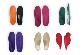
Custom orthotics can also be of benefit, especially to tighten the forefoot, alleviate metatarsalgia, correct ankles rolling in and a range of other biomechanical anomalies. These would be worn in regular footwear, when not dancing.

The biomechanics specialist podiatrists at Dubai Podiatry Centre, UK qualified and trained and led by renowned Chief Podiatrist Michelle Champlin, can be contacted on +971 4 3435390.


قلم ميشيل شامبلين

يمكن مقارنة المتطلبات الجسدية للباليه بتلك الخاصة بالعديد من الألعاب الرياضية، على الرغم من أن المتطلبات على القدمين والركبتين والوركين يمكن أن تكون شديدة. 15-20% من إصابات الرقص تصيب القدم. تميل الإصابات المزمنة إلى السيطرة لأنها ترتبط بالتأثير المتكرر لقدم الراقص على سطح صلب نسبيًا.

على عكس الرياضيين الآخرين، الذين يرتدون أحذية مصممة خصيصًا لتثبيت القدم وامتصاص الصدمات الخاصة برياضتهم (مثل التنس أو أحذية كرة الشبكة أو أحذية الرجبي)، يرتدي راقص الباليه شبشبًا رفيعًا أو حذاءًا رفيعًا فقط. لذلك، يتم امتصاص غالبية قوى التأثير عن طريق القدم والساق. تشمل العوامل التي يمكن أن تساهم في الإصابات المرتبطة بالباليه التشوهات الميكانيكية الحيوية والتقنية والتعب.

العوامل الميكانيكية الحيوية والتقنية ذات الصلة

العامل التشريحي الأكثر أهمية في الباليه الكلاسيكي هو الإقبال المناسب على الورك. تتطلب جميع الأوضاع الأساسية الخمسة أقصى قدر من الدوران الخارجي للورك. تبدأ جميع حركات الباليه أو تنتهي بأحد هذه الأوضاع. يمكن للراقصين ذوي الإقبال الأقل “الطبيعي” على الورك التعويض عن طريق فرض دوران خارجي عند مفاصل الركبة أو القدم والكاحل. التدحرج (أي ما يعادل الكبح الزائد)، هو أسلوب يستخدمه بعض الراقصين للتعويض عن دوران الورك الخارجي المحدود. نتيجة هذه المناورة هي زيادة الضغط على الهياكل الوسطى للقدم والكاحل، مما قد يؤدي إلى إصابات طويلة المدى.

يمكن أيضًا أن يسبب نوع القدم المقوسة (المقوسة العالية)، مع مفاصل منتصف الرسغ الأكثر صلابة، بعض المشكلات لراقصي الباليه. لا يؤدي تقوس القدم إلى تبديد الطاقة أيضًا، مما يعرض الهياكل الأخرى لخطر إجهاد الرباط والتهاب اللفافة وكسر الإجهاد. عندما لا يتم تقاسم جميع القوى بالتساوي بين عظام مشط القدم الخمسة، فإن مشط القدم الأكثر تحملاً للضغط يمكن أن تكون عرضة لكسور الإجهاد.

عندما تكون مقدمة القدم مفلطحة، أحيانًا بسبب انهيار القوس العرضي، يمكن أن يكون هذا مقدمة لإبهام القدم الأروح (الأورام) والأورام. الأورام شائعة في الراقصين. تميل إلى البدء في نهاية فترة المراهقة وتظهر في كل من الراقصين الذكور والإناث. يميل الألم إلى أن يكون أسوأ في نهاية الفصل.

في حين أن النطاق الطبيعي للحركة في المفصل MTP الأول هو حوالي 60 درجة عطف ظهري (أصابع القدم متجهة للأعلى) وحوالي 35 درجة ثني أخمصي (أصابع القدم متجهة للأسفل)، فإن الراقصين لديهم بشكل روتيني 90 إلى 100 درجة من العطف الظهري للسماح بالارتباط الكامل على نصف النقطة. . ويتم الحصول على ذلك عادة عن طريق الرقص بينما يتشكل الجهاز العضلي الهيكلي بحيث يمكن تشكيل المفاصل – أي خلال مرحلة الطفولة.

يمكن أن يكون التهاب السمسم والكسور السمسمانية مزعجًا للراقصين حيث قد يشفون ببطء شديد. هناك أسباب عديدة للالتهاب السمسماني، بما في ذلك الكدمات أو الكسور الإجهادية أو تنخر العظم أو التهاب المفاصل العظمي.

يمكن أيضًا أن تحدث الأعصاب المحاصرة أو المتهيجة عند الراقصين – مثل التهاب العصب أو ورم مورتون العصبي، والذي قد يحاكي التهاب السمسماني. في هذه الحالة. سيكون طبيب الأقدام الخاص بك قادرًا على التمييز بين الاثنين.

تعتبر مسامير القدم شائعة للغاية أيضًا، ويمكن استئصالها بسهولة من قبل طبيب الأقدام – لا تقم مطلقًا “بقص” تقويم العظام جراحيًا أو وضع ضمادات الذرة بحمض الساليسيليك، لأن ذلك قد يؤدي إلى تلف الجلد الصحي أو ترك ندبات.

يمكنك معرفة المزيد عن أسطح الرقص وأحذية الباليه وتأثيرها على إصابات الرقص هنا.

تقييم إصابات الباليه

يجب على الراقصين إحضار نعالهم وأحذيةهم المدببة إلى استشارة علاج الأرجل حتى يمكن فحصهم بحثًا عن علامات الإجهاد أو الاستخدام غير الطبيعي وربما حتى ارتدائها لإظهار الأوضاع التي تسبب الألم. قد يتم تمديد التقييم الميكانيكي الحيوي الكامل للقدم من قبل طبيب الأقدام ويطلب منك أداء الأوضاع المؤلمة أو المؤلمة.

علاج الإصابات: التهاب المشاش: يحتاج راقصو الباليه إلى نطاق حركة متزايد في المفصل MTP الأول (مفصل إصبع القدم الكبير). نادرًا ما يولد الأشخاص بهذا القدر من الحركة في هذا المفصل، لذلك يجب الحصول عليها عن طريق بعض قوالب صفائح النمو الموجودة في نهاية عظم إصبع القدم (الكتائب). في مرحلة المراهقة، يمكن أن يؤدي ذلك إلى الألم والالتهاب والألم أثناء النشاط الذي يخفف من الراحة. سوف تختفي الحالة بمجرد توقف الراقص عن النمو. تشبه هذه الحالة مرض أوسجود-شلاتر الذي يصيب الركبة والذي يظهر أحيانًا عند الأطفال الرياضيين.

إعادة العلاج بشكل عام، قد تكون المناورات والوضعيات التي تسبب الألم محدودة لمدة 4-5 أسابيع ثم يعاد تقديمها بحذر.

بالنسبة للأورام والوكعات، فإن الحشو بصوف الحمل اللاصق الخاص حول منطقة الألم هو وسيلة بسيطة وفعالة لتخفيف الألم. كما يستخدم أطباء الأقدام في مركز دبي لعلاج الأرجل هذه الحشوة المتخصصة لمنع تكون البثور لدى المتنزهين والعدائين.

يمكن أن تجعل فواصل أصابع القدم الهلامية بين إصبع القدم الأول والثاني القدم أكثر وظيفية، حيث تحافظ هذه المحاذاة على أقصى قدر من الحركة في MPJ. إن ارتداء أحذية واسعة وأحذية رياضية خارج صفوف الباليه سيساعد أيضًا في تعافي أصابع القدم.

على الرغم من أنه قد يُنصح بإجراء عملية جراحية (مثل استئصال الورم) للمرضى الآخرين الذين يعانون من أورام مؤلمة ومتقدمة، فإن الراقصين المحترفين يميلون إلى تجنب ذلك على الأقل خلال حياتهم المهنية النشطة لأنه عادة ما يحد من الحركة في MPJ الأولى.

لعلاج التهاب السمسماني عند الراقصين، عادةً لا يكون إجراء جراحة استئصال السمسماني ضروريًا للراقصين لأن الألم سيختفي دائمًا تقريبًا بالعلاج المحافظ وحده، على الرغم من أن هذا قد يستغرق ما يصل إلى عام. خلال هذه الفترة، قد يقوم طبيب الأقدام بإدخال وسادات صغيرة خاصة في الأحذية لتفريغ السمسمويدات ويجب على الراقصين تقليل العمل النصفي.

يمكن أن تكون أجهزة تقويم العظام المخصصة مفيدة أيضًا، خاصة لشد مقدمة القدم، وتخفيف آلام مشط القدم، وتصحيح تدحرج الكاحلين، ومجموعة من الحالات الشاذة الميكانيكية الحيوية الأخرى. سيتم ارتداؤها بالأحذية العادية عند عدم الرقص

يمكن الاتصال بأطباء الأقدام المتخصصين في الميكانيكا الحيوية في مركز دبي للعناية بالاقدام بالمملكة المتحدة، المؤهلين والمدربين بقيادة رئيسة أطباء الأقدام المشهورة ميشيل شامبلن، على 043435390


Copyright Dubai Podiatry 2026. All rights reserved.

WhatsApp us