Written by Michelle Champlin BSc Pod., M.Ch.S., S.R., Ch., (UK)
What are the ‘hamstrings’?
The hamstrings are three tendons at the back of the thighs that attach the large thigh muscle to the bone. These can be felt as the ‘strings’ on either side of the back of your knee.
Additionally, the ‘hamstring’ also includes the group of three muscles that run from beneath the pelvis along the back of the thigh, crossing behind the knee joint and ending at the lower leg. They are involved in knee flexion (bending) and hip extension.
The hamstrings play a key role in lots of everyday activities – walking, running, jumping, and controlling some movement in the trunk. In walking, they are most important in opposing the quads during knee extension.
What is a pulled hamstring?
Most hamstring injuries occur to the thick, central part of the muscle or where the muscle fibers join tendon fibers. A hamstring injury can occur if any of the tendons or muscles are stretched beyond their limit. They often occur during sudden, explosive movements, such as sprinting, lunging or jumping and are graded 1, 2 or 3 according to severity. But they can also occur more gradually, or during slower movements that overstretch your hamstring. Recurring injury is common in athletes and sportsmen, as you’re more likely to injure your hamstring if you’ve injured it previously.
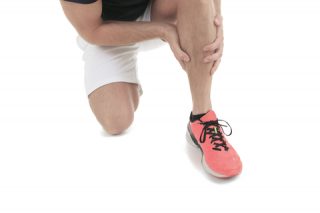
A hamstring injury is a strain or tear to the tendons or large muscles at the back of the thigh.
The length of time it takes to recover from a hamstring strain or tear will depend on how severe the injury is. A minor muscle pull or strain (grade 1) may take a few days to heal, whereas it could take weeks or months to recover from a muscle tear (grade 2 / 3).
What are the symptoms?
Mild hamstring strains (grade 1) will usually cause sudden pain and tenderness the back of your thigh. It may be painful to move your leg, but the strength of the muscle shouldn’t be affected. Partial hamstring tears (grade 2) are usually more painful and tender. There may also be some swelling or bruising at the back of the thigh and some loss of muscle strength. The most severe hamstring tears (grade 3) will usually be very painful, tender, swollen and bruised. There may have been a “popping” sensation at the time of the injury and immediate inability to weight bear on the leg.
How do injuries happen?
Hamstring injuries tend to occur due to muscle overload – either stretched beyond capacity or challenged suddenly. You might be more at risk of hamstring strain due to:
Muscle tightness: Tight muscles are vulnerable to strain. Athletes should follow a year-round program of daily stretching exercises.
Muscle imbalance: When one muscle group is much stronger than its opposing muscle group, the imbalance can lead to a strain. This frequently happens with the hamstring muscles. The quadriceps muscles at the front of the thigh are usually more powerful. During sprint work, the hamstring can tire more quickly than the quads, putting them at risk of strain.
Starting / increasing a fitness regime: If muscles are weak, they are less able to cope with the stress of exercise and are more likely to be injured.
Muscle fatigue: Tired muscles are less able to absorb energy, making them more prone to injury.
Type of Activity: Anyone can pull a hamstring, but certain athletes are more at risk:
• Athletes who participate in sports like soccer or basketball, involving bursts of running
• Runners or sprinters
• Dancers
• Older athletes whose exercise regime is primarily walking
• Adolescent athletes who are still growing.
Hamstring strains occur more often in teens because their bones and muscles grow at different rates. During a growth spurt, the child’s bones may grow faster than the muscles. The growing bone pulls the muscle tight, predisposing it to a pull or tear during a sudden jump or run.
TreatmentHamstring Injuries
Details
Written by Michelle Champlin BSc Pod., M.Ch.S., S.R., Ch., (UK)
What are the ‘hamstrings’?
The hamstrings are three tendons at the back of the thighs that attach the large thigh muscle to the bone. These can be felt as the ‘strings’ on either side of the back of your knee.
Additionally, the ‘hamstring’ also includes the group of three muscles that run from beneath the pelvis along the back of the thigh, crossing behind the knee joint and ending at the lower leg. They are involved in knee flexion (bending) and hip extension.
The hamstrings play a key role in lots of everyday activities – walking, running, jumping, and controlling some movement in the trunk. In walking, they are most important in opposing the quads during knee extension.
What is a pulled hamstring?
Most hamstring injuries occur to the thick, central part of the muscle or where the muscle fibers join tendon fibers. A hamstring injury can occur if any of the tendons or muscles are stretched beyond their limit. They often occur during sudden, explosive movements, such as sprinting, lunging or jumping and are graded 1, 2 or 3 according to severity. But they can also occur more gradually, or during slower movements that overstretch your hamstring. Recurring injury is common in athletes and sportsmen, as you’re more likely to injure your hamstring if you’ve injured it previously.
A hamstring injury is a strain or tear to the tendons or large muscles at the back of the thigh.
The length of time it takes to recover from a hamstring strain or tear will depend on how severe the injury is. A minor muscle pull or strain (grade 1) may take a few days to heal, whereas it could take weeks or months to recover from a muscle tear (grade 2 / 3).
What are the symptoms?
Mild hamstring strains (grade 1) will usually cause sudden pain and tenderness the back of your thigh. It may be painful to move your leg, but the strength of the muscle shouldn’t be affected. Partial hamstring tears (grade 2) are usually more painful and tender. There may also be some swelling or bruising at the back of the thigh and some loss of muscle strength. The most severe hamstring tears (grade 3) will usually be very painful, tender, swollen and bruised. There may have been a “popping” sensation at the time of the injury and immediate inability to weight bear on the leg.
How do injuries happen?
Hamstring injuries tend to occur due to muscle overload – either stretched beyond capacity or challenged suddenly. You might be more at risk of hamstring strain due to:
Muscle tightness: Tight muscles are vulnerable to strain. Athletes should follow a year-round program of daily stretching exercises.
Muscle imbalance: When one muscle group is much stronger than its opposing muscle group, the imbalance can lead to a strain. This frequently happens with the hamstring muscles. The quadriceps muscles at the front of the thigh are usually more powerful. During sprint work, the hamstring can tire more quickly than the quads, putting them at risk of strain.
Starting / increasing a fitness regime: If muscles are weak, they are less able to cope with the stress of exercise and are more likely to be injured.
Muscle fatigue: Tired muscles are less able to absorb energy, making them more prone to injury.
Type of Activity: Anyone can pull a hamstring, but certain athletes are more at risk:
• Athletes who participate in sports like soccer or basketball, involving bursts of running
• Runners or sprinters
• Dancers
• Older athletes whose exercise regime is primarily walking
• Adolescent athletes who are still growing.
Hamstring strains occur more often in teens because their bones and muscles grow at different rates. During a growth spurt, the child’s bones may grow faster than the muscles. The growing bone pulls the muscle tight, predisposing it to a pull or tear during a sudden jump or run.
Treatment
As with any muscle or tendon injury, immediate rest, ice, compression and elevation is important. Rest may involve the use of crutches or a brace. All but the most severe injuries heal without surgery. As pulled hamstrings are linked to poor fitness, muscle imbalance and inflexibility, avoid hamstring injuries by stepping up training regimes gradually to strengthen the hamstrings. Include hamstring strengthening exercises in balance with quad strength. Stretching daily is vital to develop flexibility and suppleness. Your Doctor or Podiatrist can guide you in stretching and strengthening rehabilitation after injury to minimize any scar tissue at the injury site or muscle atrophy. Your Podiatrist will also carry out a full lower limb biomechanical assessment to identify any contributing malalignment and advise on orthotics if required to alleviate undue stress on the hamstrings.
ما هي “أوتار الركبة”؟
أوتار الركبة عبارة عن ثلاثة أوتار في الجزء الخلفي من الفخذين تربط عضلة الفخذ الكبيرة بالعظم. يمكن الشعور بها على أنها “أوتار” على جانبي الجزء الخلفي من ركبتك.
بالإضافة إلى ذلك، تشتمل “أوتار الركبة” أيضًا على مجموعة من ثلاث عضلات تمتد من أسفل الحوض على طول الجزء الخلفي من الفخذ، وتعبر خلف مفصل الركبة وتنتهي عند أسفل الساق. إنهم يشاركون في ثني الركبة (الانحناء) وتمديد الورك.
تلعب أوتار الركبة دورًا رئيسيًا في الكثير من الأنشطة اليومية – المشي والجري والقفز والتحكم في بعض الحركة في الجذع. في المشي، هم الأكثر أهمية في معارضة الكواد أثناء تمديد الركبة.
ما هو سحب اوتار الركبة؟
تحدث معظم إصابات أوتار الركبة في الجزء المركزي السميك من العضلة أو حيث تنضم ألياف العضلات إلى ألياف الوتر. يمكن أن تحدث إصابة في أوتار الركبة إذا تم شد أي من الأوتار أو العضلات إلى ما هو أبعد من حدودها. تحدث غالبًا أثناء الحركات المفاجئة والمتفجّرة، مثل الركض السريع أو الاندفاع أو القفز، ويتم تصنيفها من 1 أو 2 أو 3 وفقًا لشدتها. ولكنها يمكن أن تحدث أيضًا بشكل تدريجي، أو أثناء الحركات البطيئة التي تزيد من إجهاد أوتار الركبة. تعد الإصابة المتكررة أمرًا شائعًا لدى الرياضيين والرياضيين، حيث من المرجح أن تصاب بأوتار الركبة إذا كنت قد أصبت بها سابقًا.
إصابة أوتار الركبة هي إجهاد أو تمزق في الأوتار أو العضلات الكبيرة في الجزء الخلفي من الفخذ.
يعتمد طول الوقت المستغرق للتعافي من إجهاد أو تمزق في أوتار الركبة على مدى خطورة الإصابة. قد يستغرق شد أو إجهاد العضلات البسيط (الدرجة 1) بضعة أيام للشفاء، في حين قد يستغرق الأمر أسابيع أو أشهر للتعافي من تمزق العضلات (الدرجة 2/3).
ما هي الاعراض؟
عادة ما تسبب سلالات أوتار الركبة الخفيفة (الدرجة 1) ألمًا مفاجئًا وألمًا في الجزء الخلفي من فخذك. قد يكون من المؤلم تحريك ساقك، ولكن لا ينبغي أن تتأثر قوة العضلات. عادة ما تكون تمزقات أوتار الركبة الجزئية (الدرجة 2) أكثر إيلامًا وإيلامًا. قد يكون هناك أيضًا بعض التورم أو الكدمات في الجزء الخلفي من الفخذ وبعض فقدان قوة العضلات. عادة ما تكون تمزقات أوتار الركبة الأكثر خطورة (الدرجة 3) مؤلمة للغاية، ومؤلمة، ومتورمة وكدمات. ربما كان هناك إحساس “بالفرقعة” وقت الإصابة وعدم القدرة المباشرة على تحمل الوزن على الساق.
كيف تحدث الإصابات؟
تميل إصابات أوتار الركبة إلى الحدوث بسبب الحمل الزائد للعضلات – إما أن تمتد إلى ما هو أبعد من طاقتها أو يتم تحديها فجأة. قد تكون أكثر عرضة لخطر الإصابة بإجهاد أوتار الركبة للأسباب التالية:
ضيق العضلات: العضلات المشدودة تكون عرضة للإجهاد. يجب على الرياضيين اتباع برنامج على مدار العام من تمارين التمدد اليومية.
اختلال توازن العضلات: عندما تكون مجموعة عضلية أقوى بكثير من مجموعة العضلات المقابلة لها، يمكن أن يؤدي عدم التوازن إلى الإجهاد. يحدث هذا بشكل متكرر مع عضلات اوتار الركبة. عادة ما تكون عضلات الفخذ الرباعية الموجودة في الجزء الأمامي من الفخذ أقوى. أثناء تمرين العدو السريع، يمكن أن تتعب أوتار الركبة بسرعة أكبر من عضلات الفخذ الرباعية، مما يعرضها لخطر الإجهاد.
البدء/زيادة نظام اللياقة البدنية: إذا كانت العضلات ضعيفة، فإنها تكون أقل قدرة على التعامل مع ضغوط التمارين الرياضية وتكون أكثر عرضة للإصابة.
إرهاق العضلات: تكون العضلات المتعبة أقل قدرة على امتصاص الطاقة، مما يجعلها أكثر عرضة للإصابة.
نوع النشاط: يمكن لأي شخص أن يسحب أوتار الركبة، ولكن بعض الرياضيين أكثر عرضة للخطر:
• الرياضيون الذين يشاركون في رياضات مثل كرة القدم أو كرة السلة، والتي تتضمن الجري السريع
• العدائين أو العدائين
• الراقصات
• الرياضيون الأكبر سناً الذين يعتمد نظام تمارينهم الرياضية على المشي بشكل أساسي
• الرياضيون المراهقون الذين ما زالوا في طور النمو.
تحدث سلالات أوتار الركبة في كثير من الأحيان عند المراهقين لأن عظامهم وعضلاتهم تنمو بمعدلات مختلفة. خلال طفرة النمو، قد تنمو عظام الطفل بشكل أسرع من العضلات. يسحب العظم النامي العضلة بقوة، مما يجعلها عرضة للشد أو التمزق أثناء القفز أو الجري المفاجئ.
علاج
كما هو الحال مع أي إصابة في العضلات أو الأوتار، من المهم الراحة الفورية والثلج والضغط والارتفاع. قد تنطوي الراحة على استخدام العكازات أو الدعامة. جميع الإصابات ما عدا الشديدة تشفى بدون جراحة. نظرًا لأن أوتار الركبة التي تم سحبها مرتبطة بضعف اللياقة البدنية وعدم توازن العضلات وعدم المرونة، تجنب إصابات أوتار الركبة عن طريق تكثيف أنظمة التدريب تدريجيًا لتقوية أوتار الركبة. قم بتضمين تمارين تقوية أوتار الركبة بالتوازن مع القوة الرباعية. يعد التمدد يوميًا أمرًا حيويًا لتطوير المرونة والليونة. يمكن لطبيبك أو طبيب الأقدام إرشادك في تمديد وتعزيز عملية إعادة التأهيل بعد الإصابة لتقليل أي أنسجة ندبية في موقع الإصابة أو ضمور العضلات. سيقوم طبيب الأقدام الخاص بك أيضًا بإجراء تقييم ميكانيكي حيوي كامل للأطراف السفلية لتحديد أي سوء استقامة مساهم وتقديم المشورة بشأن تقويم العظام إذا لزم الأمر لتخفيف الضغط غير المبرر على أوتار الركبة.